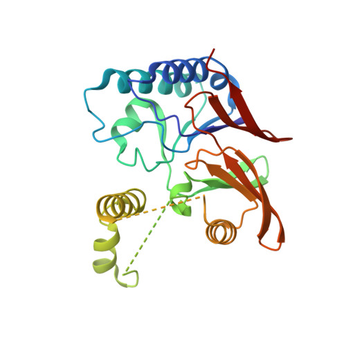
5WLY

E. coli LpxH- 8 mutations


Experimental Data Snapshot

  • Method: X-RAY DIFFRACTION
  • Resolution: 2.00 Å
  • R-Value Free: 0.227 
  • R-Value Work: 0.198 
  • R-Value Observed: 0.200 

Starting Model: experimental
View more details

wwPDB Validation   3D Report Full Report


This is version 1.5 of the entry. See complete history


Literature

The substrate-binding cap of the UDP-diacylglucosamine pyrophosphatase LpxH is highly flexible, enabling facile substrate binding and product release.

Bohl, T.E.Ieong, P.Lee, J.K.Lee, T.Kankanala, J.Shi, K.Demir, O.Kurahashi, K.Amaro, R.E.Wang, Z.Aihara, H.

(2018) J Biol Chem 293: 7969-7981

  • DOI: https://doi.org/10.1074/jbc.RA118.002503
  • Primary Citation of Related Structures:  
    5WLY

  • PubMed Abstract: 

    Gram-negative bacteria are surrounded by a secondary membrane of which the outer leaflet is composed of the glycolipid lipopolysaccharide (LPS), which guards against hydrophobic toxins, including many antibiotics. Therefore, LPS synthesis in bacteria is an attractive target for antibiotic development. LpxH is a pyrophosphatase involved in LPS synthesis, and previous structures revealed that LpxH has a helical cap that binds its lipid substrates. Here, crystallography and hydrogen-deuterium exchange MS provided evidence for a highly flexible substrate-binding cap in LpxH. Furthermore, molecular dynamics simulations disclosed how the helices of the cap may open to allow substrate entry. The predicted opening mechanism was supported by activity assays of LpxH variants. Finally, we confirmed biochemically that LpxH is inhibited by a previously identified antibacterial compound, determined the potency of this inhibitor, and modeled its binding mode in the LpxH active site. In summary, our work provides evidence that the substrate-binding cap of LpxH is highly dynamic, thus allowing for facile substrate binding and product release between the capping helices. Our results also pave the way for the rational design of more potent LpxH inhibitors.


  • Organizational Affiliation

    Department of Biochemistry, Molecular Biology & Biophysics, University of Minnesota, Twin Cities, Minneapolis, Minnesota 55455. Electronic address: bohlx031@umn.edu.


Macromolecules
Find similar proteins by:  (by identity cutoff)  |  3D Structure
Entity ID: 1
MoleculeChains Sequence LengthOrganismDetailsImage
UDP-2,3-diacylglucosamine hydrolase248Escherichia coliMutation(s): 8 
Gene Names: lpxHAUQ13_20415BK337_17315BUE81_23120EC3234A_5c00260PGD_02789
EC: 3.6.1.54
UniProt
Find proteins for P43341 (Escherichia coli (strain K12))
Explore P43341 
Go to UniProtKB:  P43341
Entity Groups  
Sequence Clusters30% Identity50% Identity70% Identity90% Identity95% Identity100% Identity
UniProt GroupP43341
Sequence Annotations
Expand
  • Reference Sequence
Experimental Data & Validation

Experimental Data

  • Method: X-RAY DIFFRACTION
  • Resolution: 2.00 Å
  • R-Value Free: 0.227 
  • R-Value Work: 0.198 
  • R-Value Observed: 0.200 
  • Space Group: P 21 2 21
Unit Cell:
Length ( Å )Angle ( ˚ )
a = 57.012α = 90
b = 62.064β = 90
c = 66.047γ = 90
Software Package:
Software NamePurpose
PHENIXrefinement
Cootmodel building
Aimlessdata scaling
PHENIXphasing
XDSdata reduction

Structure Validation

View Full Validation Report


Entry History & Funding Information

Deposition Data


Funding OrganizationLocationGrant Number
National Institutes of Health/National Institute of General Medical Sciences (NIH/NIGMS)United StatesGM118047
National Institutes of Health/National Institute of General Medical Sciences (NIH/NIGMS)United StatesP41-GM103403
Department of Energy (DOE, United States)United StatesDE-AC02-06CH11357

Revision History  (Full details and data files)

  • Version 1.0: 2018-04-11
    Type: Initial release
  • Version 1.1: 2018-04-18
    Changes: Data collection, Database references
  • Version 1.2: 2018-06-06
    Changes: Data collection, Database references, Derived calculations, Structure summary
  • Version 1.3: 2018-07-11
    Changes: Data collection, Structure summary
  • Version 1.4: 2020-01-01
    Changes: Author supporting evidence
  • Version 1.5: 2023-10-04
    Changes: Data collection, Database references, Refinement description