5WJ2 | pdb_00005wj2

Crystal structure of the green fluorescent protein Clover


Experimental Data Snapshot

  • Method: X-RAY DIFFRACTION
  • Resolution: 2.41 Å
  • R-Value Free: 
    0.217 (Depositor), 0.216 (DCC) 
  • R-Value Work: 
    0.193 (Depositor), 0.193 (DCC) 
  • R-Value Observed: 
    0.194 (Depositor) 

Starting Model: experimental
View more details

wwPDB Validation 3D Report Full Report

Validation slider image for 5WJ2

This is version 2.0 of the entry. See complete history

Literature

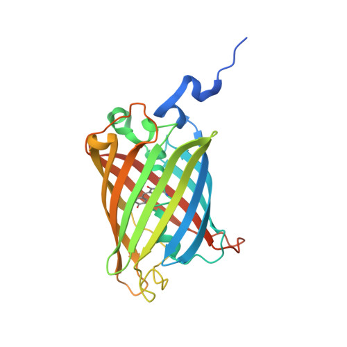
Crystal Structure of Green Fluorescent Protein Clover and Design of Clover-Based Redox Sensors.

Campbell, B.C.Petsko, G.A.Liu, C.F.

(2018) Structure 26: 225-237.e3

  • DOI: https://doi.org/10.1016/j.str.2017.12.006
  • Primary Citation Related Structures: 
    5WJ2, 5WJ3, 5WJ4

  • PubMed Abstract: 

    We have determined the crystal structure of Clover, one of the brightest fluorescent proteins, and found that its T203H/S65G mutations relative to wild-type GFP lock the critical E222 side chain in a fixed configuration that mimics the major conformer of that in EGFP. The resulting equilibrium shift to the predominantly deprotonated chromophore increases the extinction coefficient (EC), opposes photoactivation, and is responsible for the bathochromic shift. Clover's brightness can further be attributed to a π-π stacking interaction between H203 and the chromophore. Consistent with these observations, the Clover G65S mutant reversed the equilibrium shift, dramatically decreased the EC, and made Clover photoactivatable under conditions that activated photoactivatable GFP. Using the Clover structure, we rationally engineered a non-photoactivatable redox sensor, roClover1, and determined its structure as well as that of its parental template, roClover0.1. These high-resolution structures provide deeper insights into structure-function relationships in GFPs and may aid the development of excitation-improved ratiometric biosensors.


  • Organizational Affiliation
    • Helen and Robert Appel Alzheimer's Disease Research Institute and Feil Family Brain and Mind Research Institute, Weill Cornell Medicine, New York, NY 10021, USA.

Macromolecule Content 

  • Total Structure Weight: 58.45 kDa 
  • Atom Count: 3,768 
  • Modeled Residue Count: 462 
  • Deposited Residue Count: 516 
  • Unique protein chains: 1

Macromolecules

Find similar proteins by:|  3D Structure
Entity ID: 1
MoleculeChains  Sequence LengthOrganismDetailsImage
Green fluorescent protein
A, B
258Aequorea victoriaMutation(s): 11 
Gene Names: GFP
UniProt
Find proteins for P42212 (Aequorea victoria)
Explore P42212 
Go to UniProtKB:  P42212
Entity Groups
Sequence Clusters30% Identity50% Identity70% Identity90% Identity95% Identity100% Identity
UniProt GroupP42212
Sequence Annotations
Expand
Reference Sequence

Small Molecules

Modified Residues  1 Unique
IDChains TypeFormula2D DiagramParent
CR2
Query on CR2
A, B
L-PEPTIDE LINKINGC13 H13 N3 O4GLY, TYR, GLY

Experimental Data & Validation

Experimental Data

  • Method: X-RAY DIFFRACTION
  • Resolution: 2.41 Å
  • R-Value Free:  0.217 (Depositor), 0.216 (DCC) 
  • R-Value Work:  0.193 (Depositor), 0.193 (DCC) 
  • R-Value Observed: 0.194 (Depositor) 
Space Group: I 2 2 2
Unit Cell:
Length ( Å )Angle ( ˚ )
a = 53.063α = 90
b = 135.176β = 90
c = 168.589γ = 90
Software Package:
Software NamePurpose
PHENIXrefinement
HKL-2000data scaling
PDB_EXTRACTdata extraction
HKL-2000data reduction
PHASERphasing

Structure Validation

View Full Validation Report


Entry History 

Deposition Data

Revision History  (Full details and data files)

  • Version 1.0: 2018-01-17
    Type: Initial release
  • Version 1.1: 2018-01-24
    Changes: Database references
  • Version 1.2: 2018-01-31
    Changes: Database references, Source and taxonomy, Structure summary
  • Version 1.3: 2018-02-14
    Changes: Database references
  • Version 1.4: 2023-10-04
    Changes: Data collection, Database references, Refinement description
  • Version 1.5: 2023-11-15
    Changes: Data collection
  • Version 1.6: 2024-10-09
    Changes: Structure summary
  • Version 2.0: 2026-03-18
    Changes: Polymer sequence