5WD8

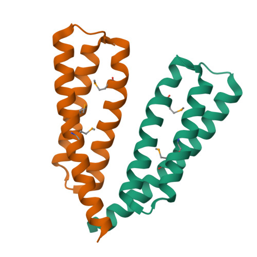
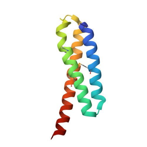
Crystal structure of Legionella pneumophila effector lpg2328


Experimental Data Snapshot

  • Method: X-RAY DIFFRACTION
  • Resolution: 1.94 Å
  • R-Value Free: 0.270 
  • R-Value Work: 0.216 
  • R-Value Observed: 0.219 

wwPDB Validation   3D Report Full Report


This is version 1.4 of the entry. See complete history


Literature

Crystal structure of the Legionella effector Lem22.

Kozlov, G.Wong, K.Gehring, K.

(2018) Proteins 86: 263-267

  • DOI: https://doi.org/10.1002/prot.25427
  • Primary Citation of Related Structures:  
    5WD8, 5WD9

  • PubMed Abstract: 

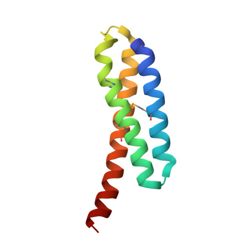
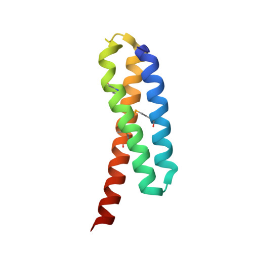
    Legionella pneumophila is a pathogen causing severe pneumonia in humans called Legionnaires' disease. Lem22 is a previously uncharacterized effector protein conserved in multiple Legionella strains. Here, we report the crystal structure of Lem22 from the Philadelphia strain, also known as lpg2328, at 1.40 Å resolution. The structure shows an up-and-down three-helical bundle with a significant structural similarity to a number of protein-binding domains involved in apoptosis and membrane trafficking. Sequence conservation identifies a putative functional site on the interface of helices 2 and 3. The structure is an important step toward a functional characterization of Lem22.


  • Organizational Affiliation

    Department of Biochemistry, Groupe de recherche axé sur la structure des protéines, McGill University, Montreal, QC, H3G 0B1, Canada.


Macromolecules
Find similar proteins by:  (by identity cutoff)  |  3D Structure
Entity ID: 1
MoleculeChains Sequence LengthOrganismDetailsImage
Lem22
A, B
91Legionella pneumophila subsp. pneumophila ATCC 43290Mutation(s): 0 
Gene Names: lp12_2320
Entity Groups  
Sequence Clusters30% Identity50% Identity70% Identity90% Identity95% Identity100% Identity
Sequence Annotations
Expand
  • Reference Sequence
Small Molecules
Modified Residues  1 Unique
IDChains TypeFormula2D DiagramParent
MSE
Query on MSE
A, B
L-PEPTIDE LINKINGC5 H11 N O2 SeMET
Experimental Data & Validation

Experimental Data

  • Method: X-RAY DIFFRACTION
  • Resolution: 1.94 Å
  • R-Value Free: 0.270 
  • R-Value Work: 0.216 
  • R-Value Observed: 0.219 
  • Space Group: P 1 21 1
Unit Cell:
Length ( Å )Angle ( ˚ )
a = 23.044α = 90
b = 81.143β = 90.81
c = 45.684γ = 90
Software Package:
Software NamePurpose
PHENIXrefinement
HKL-2000data reduction
HKL-2000data scaling
PHENIXphasing

Structure Validation

View Full Validation Report


Entry History & Funding Information

Deposition Data


Funding OrganizationLocationGrant Number
Canadian Institutes of Health Research (CIHR)Canada--

Revision History  (Full details and data files)

  • Version 1.0: 2017-11-29
    Type: Initial release
  • Version 1.1: 2017-12-13
    Changes: Database references, Structure summary
  • Version 1.2: 2018-04-25
    Changes: Data collection, Database references
  • Version 1.3: 2020-01-08
    Changes: Author supporting evidence
  • Version 1.4: 2024-10-30
    Changes: Data collection, Database references, Structure summary