5WC1 | pdb_00005wc1

katanin AAA ATPase domain


Experimental Data Snapshot

  • Method: X-RAY DIFFRACTION
  • Resolution: 3.30 Å
  • R-Value Free: 
    0.307 (Depositor), 0.306 (DCC) 
  • R-Value Work: 
    0.247 (Depositor), 0.254 (DCC) 
  • R-Value Observed: 
    0.254 (Depositor) 

Starting Model: experimental
View more details

wwPDB Validation 3D Report Full Report

Validation slider image for 5WC1

This is version 1.3 of the entry. See complete history

Literature

Katanin spiral and ring structures shed light on power stroke for microtubule severing.

Zehr, E.Szyk, A.Piszczek, G.Szczesna, E.Zuo, X.Roll-Mecak, A.

(2017) Nat Struct Mol Biol 24: 717-725

  • DOI: https://doi.org/10.1038/nsmb.3448
  • Primary Citation Related Structures: 
    5WC0, 5WC1, 5WCB

  • PubMed Abstract: 

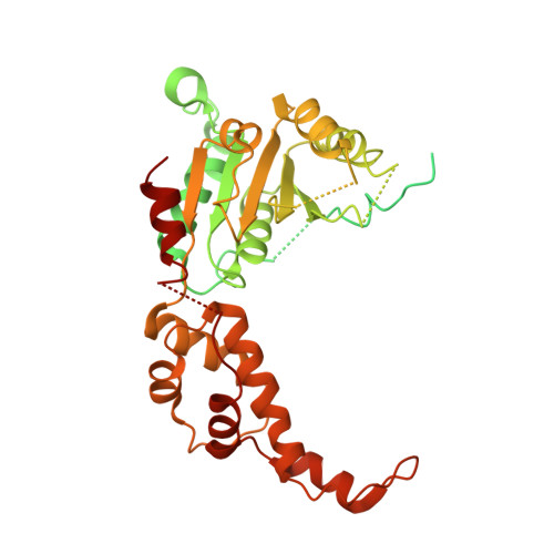
    Microtubule-severing enzymes katanin, spastin and fidgetin are AAA ATPases important for the biogenesis and maintenance of complex microtubule arrays in axons, spindles and cilia. Because of a lack of known 3D structures for these enzymes, their mechanism of action has remained poorly understood. Here we report the X-ray crystal structure of the monomeric AAA katanin module from Caenorhabditis elegans and cryo-EM reconstructions of the hexamer in two conformations. The structures reveal an unexpected asymmetric arrangement of the AAA domains mediated by structural elements unique to microtubule-severing enzymes and critical for their function. The reconstructions show that katanin cycles between open spiral and closed ring conformations, depending on the ATP occupancy of a gating protomer that tenses or relaxes interprotomer interfaces. Cycling of the hexamer between these conformations would provide the power stroke for microtubule severing.


  • Organizational Affiliation
    • Cell Biology and Biophysics Unit, Porter Neuroscience Research Center, National Institute of Neurological Disorders and Stroke, Bethesda, Maryland, USA.

Macromolecule Content 

  • Total Structure Weight: 51.99 kDa 
  • Atom Count: 1,792 
  • Modeled Residue Count: 262 
  • Deposited Residue Count: 472 
  • Unique protein chains: 1

Macromolecules

Find similar proteins by:|  3D Structure
Entity ID: 1
MoleculeChains  Sequence LengthOrganismDetailsImage
Meiotic spindle formation protein mei-1472Caenorhabditis elegansMutation(s): 1 
Gene Names: mei-1T01G9.5
EC: 3.6.4.3 (PDB Primary Data), 5.6.1.1 (UniProt)
UniProt
Find proteins for P34808 (Caenorhabditis elegans)
Explore P34808 
Go to UniProtKB:  P34808
Entity Groups
Sequence Clusters30% Identity50% Identity70% Identity90% Identity95% Identity100% Identity
UniProt GroupP34808
Sequence Annotations
Expand
Reference Sequence

Experimental Data & Validation

Experimental Data

  • Method: X-RAY DIFFRACTION
  • Resolution: 3.30 Å
  • R-Value Free:  0.307 (Depositor), 0.306 (DCC) 
  • R-Value Work:  0.247 (Depositor), 0.254 (DCC) 
  • R-Value Observed: 0.254 (Depositor) 
Space Group: P 65
Unit Cell:
Length ( Å )Angle ( ˚ )
a = 99.363α = 90
b = 99.363β = 90
c = 76.581γ = 120
Software Package:
Software NamePurpose
PHENIXrefinement
HKL-2000data reduction
SCALEPACKdata scaling
PHENIXphasing

Structure Validation

View Full Validation Report


Entry History 

Deposition Data

Revision History  (Full details and data files)

  • Version 1.0: 2017-08-09
    Type: Initial release
  • Version 1.1: 2017-08-23
    Changes: Database references
  • Version 1.2: 2017-09-20
    Changes: Database references
  • Version 1.3: 2023-10-04
    Changes: Data collection, Database references, Refinement description