5WB2 | pdb_00005wb2

US28 bound to engineered chemokine CX3CL1.35 and nanobodies


Experimental Data Snapshot

  • Method: X-RAY DIFFRACTION
  • Resolution: 3.50 Å
  • R-Value Free: 
    0.296 (Depositor), 0.301 (DCC) 
  • R-Value Work: 
    0.252 (Depositor), 0.257 (DCC) 
  • R-Value Observed: 
    0.256 (Depositor) 

Starting Model: experimental
View more details

wwPDB Validation 3D Report Full Report

Validation slider image for 5WB2

Ligand Structure Quality Assessment 


This is version 2.1 of the entry. See complete history

Literature

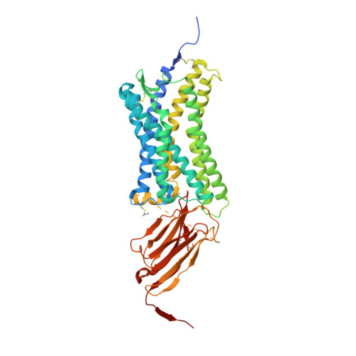
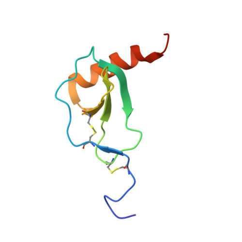
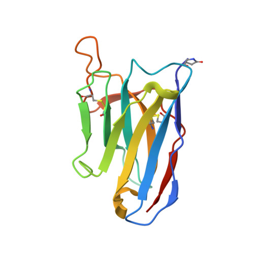
Viral GPCR US28 can signal in response to chemokine agonists of nearly unlimited structural degeneracy.

Miles, T.F.Spiess, K.Jude, K.M.Tsutsumi, N.Burg, J.S.Ingram, J.R.Waghray, D.Hjorto, G.M.Larsen, O.Ploegh, H.L.Rosenkilde, M.M.Garcia, K.C.

(2018) Elife 7

  • DOI: https://doi.org/10.7554/eLife.35850
  • Primary Citation Related Structures: 
    5WB1, 5WB2

  • PubMed Abstract: 

    Human cytomegalovirus has hijacked and evolved a human G-protein-coupled receptor into US28, which functions as a promiscuous chemokine 'sink' to facilitate evasion of host immune responses. To probe the molecular basis of US28's unique ligand cross-reactivity, we deep-sequenced CX3CL1 chemokine libraries selected on 'molecular casts' of the US28 active-state and find that US28 can engage thousands of distinct chemokine sequences, many of which elicit diverse signaling outcomes. The structure of a G-protein-biased CX3CL1-variant in complex with US28 revealed an entirely unique chemokine amino terminal peptide conformation and remodeled constellation of receptor-ligand interactions. Receptor signaling, however, is remarkably robust to mutational disruption of these interactions. Thus, US28 accommodates and functionally discriminates amongst highly degenerate chemokine sequences by sensing the steric bulk of the ligands, which distort both receptor extracellular loops and the walls of the ligand binding pocket to varying degrees, rather than requiring sequence-specific bonding chemistries for recognition and signaling.


  • Organizational Affiliation
    • Department of Molecular and Cellular Physiology, Stanford University School of Medicine, Stanford, United States.

Macromolecule Content 

  • Total Structure Weight: 77.4 kDa 
  • Atom Count: 4,907 
  • Modeled Residue Count: 621 
  • Deposited Residue Count: 667 
  • Unique protein chains: 3

Macromolecules

Find similar proteins by:|  3D Structure
Entity ID: 1
MoleculeChains  Sequence LengthOrganismDetailsImage
Envelope protein US28, nanobody 7 fusion protein460Human betaherpesvirus 5Lama glama
This entity is chimeric
Mutation(s): 0 
Gene Names: US28
Membrane Entity: Yes 
UniProt
Find proteins for P69333 (Human cytomegalovirus (strain Towne))
Explore P69333 
Go to UniProtKB:  P69333
Entity Groups
Sequence Clusters30% Identity50% Identity70% Identity90% Identity95% Identity100% Identity
UniProt GroupP69333
Sequence Annotations
Expand
Reference Sequence
Find similar proteins by:|  3D Structure
Entity ID: 2
MoleculeChains  Sequence LengthOrganismDetailsImage
CX3CL1 protein76Homo sapiensMutation(s): 0 
Gene Names: CX3CL1
Membrane Entity: Yes 
UniProt & NIH Common Fund Data Resources
Find proteins for P78423 (Homo sapiens)
Explore P78423 
Go to UniProtKB:  P78423
PHAROS:  P78423
GTEx:  ENSG00000006210 
Entity Groups
Sequence Clusters30% Identity50% Identity70% Identity90% Identity95% Identity100% Identity
UniProt GroupP78423
Sequence Annotations
Expand
Reference Sequence
Find similar proteins by:|  3D Structure
Entity ID: 3
MoleculeChains  Sequence LengthOrganismDetailsImage
nanobody B1C [auth D]131synthetic constructMutation(s): 0 

Small Molecules

Ligands 4 Unique
IDChains Name / Formula / InChI Key2D Diagram3D Interactions
CLR

Query on CLR


Download:Ideal Coordinates CCD File
D [auth A],
E [auth A]
CHOLESTEROL
C27 H46 O
HVYWMOMLDIMFJA-DPAQBDIFSA-N
OLC

Query on OLC


Download:Ideal Coordinates CCD File
F [auth A],
G [auth A],
H [auth A],
I [auth A]
(2R)-2,3-dihydroxypropyl (9Z)-octadec-9-enoate
C21 H40 O4
RZRNAYUHWVFMIP-GDCKJWNLSA-N
MES

Query on MES


Download:Ideal Coordinates CCD File
K [auth B]2-(N-MORPHOLINO)-ETHANESULFONIC ACID
C6 H13 N O4 S
SXGZJKUKBWWHRA-UHFFFAOYSA-N
ZN

Query on ZN


Download:Ideal Coordinates CCD File
J [auth B]ZINC ION
Zn
PTFCDOFLOPIGGS-UHFFFAOYSA-N
Modified Residues  2 Unique
IDChains TypeFormula2D DiagramParent
YCM
Query on YCM
A
L-PEPTIDE LINKINGC5 H10 N2 O3 SCYS
PCA
Query on PCA
C [auth D]L-PEPTIDE LINKINGC5 H7 N O3GLN

Experimental Data & Validation

Experimental Data

  • Method: X-RAY DIFFRACTION
  • Resolution: 3.50 Å
  • R-Value Free:  0.296 (Depositor), 0.301 (DCC) 
  • R-Value Work:  0.252 (Depositor), 0.257 (DCC) 
  • R-Value Observed: 0.256 (Depositor) 
Space Group: P 21 21 2
Unit Cell:
Length ( Å )Angle ( ˚ )
a = 47.17α = 90
b = 128.94β = 90
c = 127.74γ = 90
Software Package:
Software NamePurpose
PHENIXrefinement
XDSdata reduction
XSCALEdata scaling
PHASERphasing

Structure Validation

View Full Validation Report


Ligand Structure Quality Assessment 


Entry History 

& Funding Information

Deposition Data


Funding OrganizationLocationGrant Number
National Institutes of Health/National Institute of General Medical Sciences (NIH/NIGMS)United StatesGM097015

Revision History  (Full details and data files)

  • Version 1.0: 2018-06-13
    Type: Initial release
  • Version 1.1: 2018-12-26
    Changes: Data collection, Database references
  • Version 2.0: 2020-01-01
    Changes: Author supporting evidence, Polymer sequence
  • Version 2.1: 2023-10-04
    Changes: Data collection, Database references, Derived calculations, Refinement description