5WB0 | pdb_00005wb0

Crystal structure of human metapneumovirus fusion glycoprotein stabilized in the prefusion state

  • Classification: VIRAL PROTEIN
  • Organism(s): human metapneumovirus
  • Expression System: Chlorocebus aethiops
  • Mutation(s): Yes 

  • Deposited: 2017-06-27 Released: 2017-11-22 
  • Deposition Author(s): Battles, M.B., McLellan, J.S.
  • Funding Organization(s): National Institutes of Health/National Institute Of Allergy and Infectious Diseases (NIH/NIAID), National Institutes of Health/National Institute of General Medical Sciences (NIH/NIGMS)

Experimental Data Snapshot

  • Method: X-RAY DIFFRACTION
  • Resolution: 2.60 Å
  • R-Value Free: 
    0.207 (Depositor), 0.207 (DCC) 
  • R-Value Work: 
    0.170 (Depositor), 0.169 (DCC) 
  • R-Value Observed: 
    0.172 (Depositor) 

Starting Models: experimental
View more details

wwPDB Validation 3D Report Full Report

Validation slider image for 5WB0

Ligand Structure Quality Assessment 


This is version 2.2 of the entry. See complete history

Literature

Structure and immunogenicity of pre-fusion-stabilized human metapneumovirus F glycoprotein.

Battles, M.B.Mas, V.Olmedillas, E.Cano, O.Vazquez, M.Rodriguez, L.Melero, J.A.McLellan, J.S.

(2017) Nat Commun 8: 1528-1528

  • DOI: https://doi.org/10.1038/s41467-017-01708-9
  • Primary Citation Related Structures: 
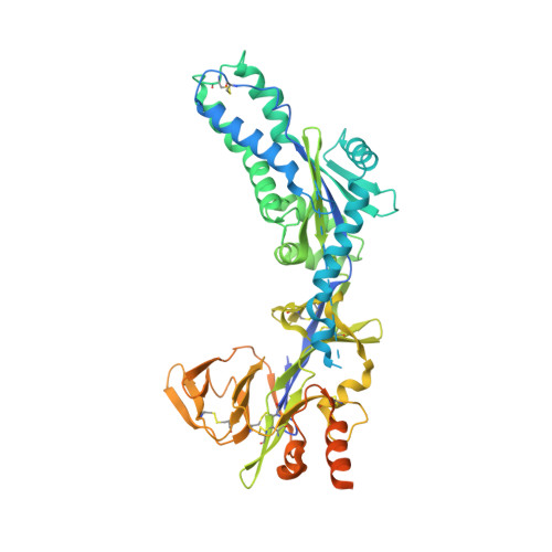
    5WB0

  • PubMed Abstract: 

    Human metapneumovirus (hMPV) is a frequent cause of bronchiolitis in young children. Its F glycoprotein mediates virus-cell membrane fusion and is the primary target of neutralizing antibodies. The inability to produce recombinant hMPV F glycoprotein in the metastable pre-fusion conformation has hindered structural and immunological studies. Here, we engineer a pre-fusion-stabilized hMPV F ectodomain and determine its crystal structure to 2.6 Å resolution. This structure reveals molecular determinants of strain-dependent acid-induced fusion, as well as insights into refolding from pre- to post-fusion conformations. A dense glycan shield at the apex of pre-fusion hMPV F suggests that antibodies against this site may not be elicited by host immune responses, which is confirmed by depletion studies of human immunoglobulins and by mouse immunizations. This is a major difference with pre-fusion F from human respiratory syncytial virus (hRSV), and collectively our results should facilitate development of effective hMPV vaccine candidates.


  • Organizational Affiliation
    • Department of Biochemistry and Cell Biology, Geisel School of Medicine at Dartmouth, Hanover, NH, 03755, USA.

Macromolecule Content 

  • Total Structure Weight: 61.11 kDa 
  • Atom Count: 3,602 
  • Modeled Residue Count: 442 
  • Deposited Residue Count: 542 
  • Unique protein chains: 1

Macromolecules

Find similar proteins by:|  3D Structure
Entity ID: 1
MoleculeChains  Sequence LengthOrganismDetailsImage
Fusion glycoprotein F0A [auth F]542human metapneumovirusMutation(s): 3 
UniProt
Find proteins for Q1A2Z0 (human metapneumovirus)
Explore Q1A2Z0 
Go to UniProtKB:  Q1A2Z0
Entity Groups
Sequence Clusters30% Identity50% Identity70% Identity90% Identity95% Identity100% Identity
UniProt GroupQ1A2Z0
Glycosylation
Glycosylation Sites: 3
Sequence Annotations
Expand
Reference Sequence

Oligosaccharides

Help  
Entity ID: 2
MoleculeChains Length2D Diagram GlycosylationD Interactions
2-acetamido-2-deoxy-beta-D-glucopyranose-(1-4)-2-acetamido-2-deoxy-beta-D-glucopyranoseB [auth A]2N-Glycosylation
Glycosylation Resources
GlyTouCan: G42666HT
GlyCosmos: G42666HT
GlyGen: G42666HT
Entity ID: 3
MoleculeChains Length2D Diagram GlycosylationD Interactions
alpha-D-mannopyranose-(1-3)-beta-D-mannopyranose-(1-4)-2-acetamido-2-deoxy-beta-D-glucopyranose-(1-4)-2-acetamido-2-deoxy-beta-D-glucopyranoseC [auth B]4N-Glycosylation
Glycosylation Resources
GlyTouCan: G81315DD
GlyCosmos: G81315DD
GlyGen: G81315DD

Experimental Data & Validation

Experimental Data

  • Method: X-RAY DIFFRACTION
  • Resolution: 2.60 Å
  • R-Value Free:  0.207 (Depositor), 0.207 (DCC) 
  • R-Value Work:  0.170 (Depositor), 0.169 (DCC) 
  • R-Value Observed: 0.172 (Depositor) 
Space Group: I 21 3
Unit Cell:
Length ( Å )Angle ( ˚ )
a = 177.7α = 90
b = 177.7β = 90
c = 177.7γ = 90
Software Package:
Software NamePurpose
PHENIXrefinement
MOSFLMdata reduction
Aimlessdata scaling
PDB_EXTRACTdata extraction
PHASERphasing

Structure Validation

View Full Validation Report


Ligand Structure Quality Assessment 


Entry History 

& Funding Information

Deposition Data


Funding OrganizationLocationGrant Number
National Institutes of Health/National Institute Of Allergy and Infectious Diseases (NIH/NIAID)United States5T32AI007519-18
National Institutes of Health/National Institute of General Medical Sciences (NIH/NIGMS)United StatesP20GM113132

Revision History  (Full details and data files)

  • Version 1.0: 2017-11-22
    Type: Initial release
  • Version 1.1: 2017-12-06
    Changes: Author supporting evidence
  • Version 1.2: 2018-04-04
    Changes: Data collection, Database references
  • Version 1.3: 2019-12-11
    Changes: Author supporting evidence
  • Version 2.0: 2020-07-29
    Type: Remediation
    Reason: Carbohydrate remediation
    Changes: Advisory, Atomic model, Data collection, Derived calculations, Structure summary
  • Version 2.1: 2023-10-04
    Changes: Data collection, Database references, Derived calculations, Refinement description, Structure summary
  • Version 2.2: 2024-11-13
    Changes: Structure summary