5WAN | pdb_00005wan

Crystal Structure of a flavoenzyme RutA in the pyrimidine catabolic pathway


Experimental Data Snapshot

  • Method: X-RAY DIFFRACTION
  • Resolution: 1.80 Å
  • R-Value Free: 
    0.172 (Depositor), 0.173 (DCC) 
  • R-Value Work: 
    0.147 (Depositor), 0.148 (DCC) 
  • R-Value Observed: 
    0.149 (Depositor) 

Starting Models: experimental
View more details

wwPDB Validation 3D Report Full Report

Validation slider image for 5WAN

Ligand Structure Quality Assessment 


This is version 1.1 of the entry. See complete history

Literature

Catalysis of a flavoenzyme-mediated amide hydrolysis.

Mukherjee, T.Zhang, Y.Abdelwahed, S.Ealick, S.E.Begley, T.P.

(2010) J Am Chem Soc 132: 5550-1

  • DOI: https://doi.org/10.1021/ja9107676
  • Primary Citation Related Structures: 
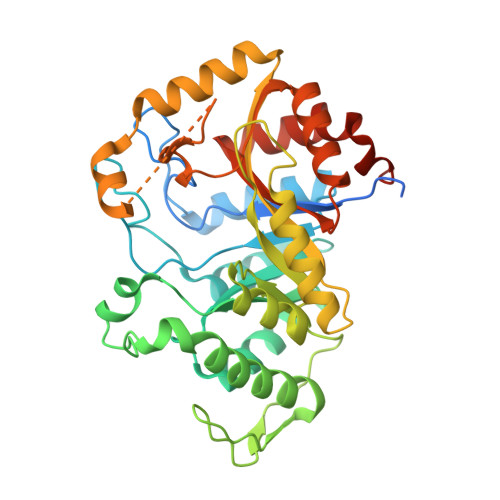
    5WAN

  • PubMed Abstract: 

    A new pyrimidine catabolic pathway (the Rut pathway) was recently discovered in Escherichia coli K12. In this pathway, uracil is converted to 3-hydroxypropionate, ammonia, and carbon dioxide. The seven-gene Rut operon is required for this conversion. Here we demonstrate that the flavoenzyme RutA catalyzes the initial uracil ring-opening reaction to give 3-ureidoacrylate. This reaction, while formally a hydrolysis reaction, proceeds by an oxidative mechanism initiated by the addition of a flavin hydroperoxide to the C4 carbonyl. While peroxide-catalyzed amide hydrolysis has chemical precedent, we are not aware of a prior example of analogous chemistry catalyzed by flavin hydroperoxides. This study further illustrates the extraordinary catalytic versatility of the flavin cofactor.


  • Organizational Affiliation
    • Department of Chemistry and Chemical Biology, Cornell University, Ithaca, New York 14853, USA.

Macromolecule Content 

  • Total Structure Weight: 45.83 kDa 
  • Atom Count: 3,150 
  • Modeled Residue Count: 341 
  • Deposited Residue Count: 403 
  • Unique protein chains: 1

Macromolecules

Find similar proteins by:|  3D Structure
Entity ID: 1
MoleculeChains  Sequence LengthOrganismDetailsImage
Pyrimidine monooxygenase RutA403Escherichia coliMutation(s): 0 
Gene Names: rutABUE81_22975BW690_24255
EC: 1.14.99.46
UniProt
Find proteins for P75898 (Escherichia coli (strain K12))
Explore P75898 
Go to UniProtKB:  P75898
Entity Groups
Sequence Clusters30% Identity50% Identity70% Identity90% Identity95% Identity100% Identity
UniProt GroupP75898
Sequence Annotations
Expand
Reference Sequence

Small Molecules

Ligands 4 Unique
IDChains Name / Formula / InChI Key2D Diagram3D Interactions
FMN

Query on FMN


Download:Ideal Coordinates CCD File
J [auth A]FLAVIN MONONUCLEOTIDE
C17 H21 N4 O9 P
FVTCRASFADXXNN-SCRDCRAPSA-N
URA

Query on URA


Download:Ideal Coordinates CCD File
B [auth A]URACIL
C4 H4 N2 O2
ISAKRJDGNUQOIC-UHFFFAOYSA-N
SO4

Query on SO4


Download:Ideal Coordinates CCD File
C [auth A],
D [auth A],
E [auth A],
F [auth A]
SULFATE ION
O4 S
QAOWNCQODCNURD-UHFFFAOYSA-L
GOL

Query on GOL


Download:Ideal Coordinates CCD File
G [auth A],
H [auth A],
I [auth A]
GLYCEROL
C3 H8 O3
PEDCQBHIVMGVHV-UHFFFAOYSA-N

Experimental Data & Validation

Experimental Data

  • Method: X-RAY DIFFRACTION
  • Resolution: 1.80 Å
  • R-Value Free:  0.172 (Depositor), 0.173 (DCC) 
  • R-Value Work:  0.147 (Depositor), 0.148 (DCC) 
  • R-Value Observed: 0.149 (Depositor) 
Space Group: P 31 2 1
Unit Cell:
Length ( Å )Angle ( ˚ )
a = 87.85α = 90
b = 87.85β = 90
c = 96.368γ = 120
Software Package:
Software NamePurpose
PHENIXrefinement
HKL-2000data collection
HKL-2000data scaling
PDB_EXTRACTdata extraction
MrBUMPphasing
HKL-2000data reduction

Structure Validation

View Full Validation Report


Ligand Structure Quality Assessment 


Entry History 

Deposition Data

Revision History  (Full details and data files)

  • Version 1.0: 2017-07-19
    Type: Initial release
  • Version 1.1: 2023-10-04
    Changes: Data collection, Database references, Refinement description