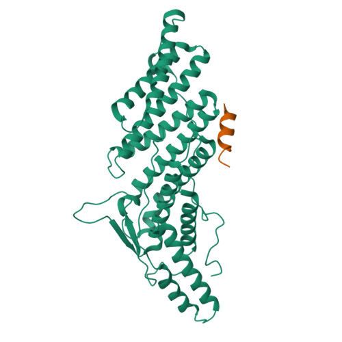
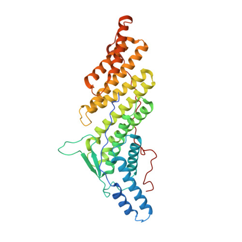
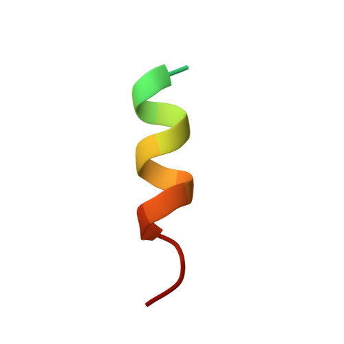
5WA1

CHMP4C A232T in complex with ALIX BRO1


Experimental Data Snapshot

  • Method: X-RAY DIFFRACTION
  • Resolution: 1.87 Å
  • R-Value Free: 0.215 
  • R-Value Work: 0.194 
  • R-Value Observed: 0.195 

Starting Model: experimental
View more details

wwPDB Validation   3D Report Full Report


This is version 1.3 of the entry. See complete history


Literature

A cancer-associated polymorphism in ESCRT-III disrupts the abscission checkpoint and promotes genome instability.

Sadler, J.B.A.Wenzel, D.M.Williams, L.K.Guindo-Martinez, M.Alam, S.L.Mercader, J.M.Torrents, D.Ullman, K.S.Sundquist, W.I.Martin-Serrano, J.

(2018) Proc Natl Acad Sci U S A 115: E8900-E8908

  • DOI: https://doi.org/10.1073/pnas.1805504115
  • Primary Citation of Related Structures:  
    5V3R, 5WA1

  • PubMed Abstract: 

    Cytokinetic abscission facilitates the irreversible separation of daughter cells. This process requires the endosomal-sorting complexes required for transport (ESCRT) machinery and is tightly regulated by charged multivesicular body protein 4C (CHMP4C), an ESCRT-III subunit that engages the abscission checkpoint (NoCut) in response to mitotic problems such as persisting chromatin bridges within the midbody. Importantly, a human polymorphism in CHMP4C (rs35094336, CHMP4C T232 ) increases cancer susceptibility. Here, we explain the structural and functional basis for this cancer association: The CHMP4C T232 allele unwinds the C-terminal helix of CHMP4C, impairs binding to the early-acting ESCRT factor ALIX, and disrupts the abscission checkpoint. Cells expressing CHMP4C T232 exhibit increased levels of DNA damage and are sensitized to several conditions that increase chromosome missegregation, including DNA replication stress, inhibition of the mitotic checkpoint, and loss of p53. Our data demonstrate the biological importance of the abscission checkpoint and suggest that dysregulation of abscission by CHMP4C T232 may synergize with oncogene-induced mitotic stress to promote genomic instability and tumorigenesis.


  • Organizational Affiliation

    Department of Infectious Diseases, Faculty of Life Sciences and Medicine, King's College London, SE1 9RT London, United Kingdom.


Macromolecules
Find similar proteins by:  (by identity cutoff)  |  3D Structure
Entity ID: 1
MoleculeChains Sequence LengthOrganismDetailsImage
Programmed cell death 6-interacting protein379Homo sapiensMutation(s): 0 
Gene Names: PDCD6IPAIP1ALIXKIAA1375
UniProt & NIH Common Fund Data Resources
Find proteins for Q8WUM4 (Homo sapiens)
Explore Q8WUM4 
Go to UniProtKB:  Q8WUM4
PHAROS:  Q8WUM4
GTEx:  ENSG00000170248 
Entity Groups  
Sequence Clusters30% Identity50% Identity70% Identity90% Identity95% Identity100% Identity
UniProt GroupQ8WUM4
Sequence Annotations
Expand
  • Reference Sequence

Find similar proteins by:  Sequence   |   3D Structure  

Entity ID: 2
MoleculeChains Sequence LengthOrganismDetailsImage
Charged multivesicular body protein 4c18Homo sapiensMutation(s): 1 
UniProt & NIH Common Fund Data Resources
Find proteins for Q96CF2 (Homo sapiens)
Explore Q96CF2 
Go to UniProtKB:  Q96CF2
PHAROS:  Q96CF2
GTEx:  ENSG00000164695 
Entity Groups  
Sequence Clusters30% Identity50% Identity70% Identity90% Identity95% Identity100% Identity
UniProt GroupQ96CF2
Sequence Annotations
Expand
  • Reference Sequence
Experimental Data & Validation

Experimental Data

  • Method: X-RAY DIFFRACTION
  • Resolution: 1.87 Å
  • R-Value Free: 0.215 
  • R-Value Work: 0.194 
  • R-Value Observed: 0.195 
  • Space Group: C 1 2 1
Unit Cell:
Length ( Å )Angle ( ˚ )
a = 120.979α = 90
b = 61.45β = 121.7
c = 76.187γ = 90
Software Package:
Software NamePurpose
PHENIXrefinement
XSCALEdata scaling
PDB_EXTRACTdata extraction
XDSdata reduction
PHASERphasing

Structure Validation

View Full Validation Report


Entry History & Funding Information

Deposition Data


Funding OrganizationLocationGrant Number
National Institutes of Health/National Institute of General Medical Sciences (NIH/NIGMS)United StatesR01 GM112080

Revision History  (Full details and data files)

  • Version 1.0: 2018-09-19
    Type: Initial release
  • Version 1.1: 2018-10-03
    Changes: Data collection, Database references
  • Version 1.2: 2020-01-01
    Changes: Author supporting evidence
  • Version 1.3: 2023-10-04
    Changes: Data collection, Database references, Refinement description