5W3X | pdb_00005w3x

Crystal structure of PopP2 in complex with IP6, AcCoA and the WRKY domain of RRS1-R .


Experimental Data Snapshot

  • Method: X-RAY DIFFRACTION
  • Resolution: 2.00 Å
  • R-Value Free: 
    0.212 (Depositor), 0.214 (DCC) 
  • R-Value Work: 
    0.183 (Depositor), 0.188 (DCC) 
  • R-Value Observed: 
    0.184 (Depositor) 

Starting Model: experimental
View more details

wwPDB Validation 3D Report Full Report

Validation slider image for 5W3X

Ligand Structure Quality Assessment 


This is version 1.4 of the entry. See complete history

Literature

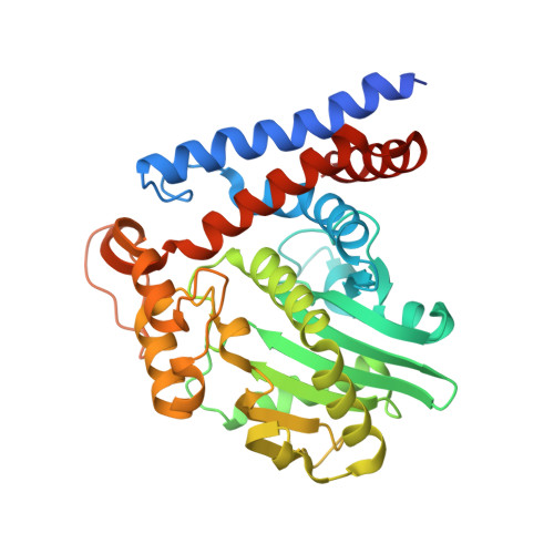
Mechanism of host substrate acetylation by a YopJ family effector.

Zhang, Z.M.Ma, K.W.Gao, L.Hu, Z.Schwizer, S.Ma, W.Song, J.

(2017) Nat Plants 3: 17115-17115

  • DOI: https://doi.org/10.1038/nplants.2017.115
  • Primary Citation Related Structures: 
    5W3T, 5W3X, 5W3Y, 5W40

  • PubMed Abstract: 

    The Yersinia outer protein J (YopJ) family of bacterial effectors depends on a novel acetyltransferase domain to acetylate signalling proteins from plant and animal hosts. However, the underlying mechanism is unclear. Here, we report the crystal structures of PopP2, a YopJ effector produced by the plant pathogen Ralstonia solanacearum, in complex with inositol hexaphosphate (InsP 6 ), acetyl-coenzyme A (AcCoA) and/or substrate Resistance to Ralstonia solanacearum 1 (RRS1-R) WRKY . PopP2 recognizes the WRKYGQK motif of RRS1-R WRKY to position a targeted lysine in the active site for acetylation. Importantly, the PopP2-RRS1-R WRKY association is allosterically regulated by InsP 6 binding, suggesting a previously unidentified role of the eukaryote-specific cofactor in substrate interaction. Furthermore, we provide evidence for the reaction intermediate of PopP2-mediated acetylation, an acetyl-cysteine covalent adduct, lending direct support to the 'ping-pong'-like catalytic mechanism proposed for YopJ effectors. Our study provides critical mechanistic insights into the virulence activity of YopJ class of acetyltransferases.


  • Organizational Affiliation
    • Department of Biochemistry, University of California, Riverside, California 92521, USA.

Macromolecule Content 

  • Total Structure Weight: 99.15 kDa 
  • Atom Count: 6,934 
  • Modeled Residue Count: 817 
  • Deposited Residue Count: 864 
  • Unique protein chains: 2

Macromolecules

Find similar proteins by:|  3D Structure
Entity ID: 1
MoleculeChains  Sequence LengthOrganismDetailsImage
PopP2 protein
A, C
352Ralstonia solanacearumMutation(s): 0 
Gene Names: poppRUN1985_v1_1190012
UniProt
Find proteins for A0A0S4VB05 (Ralstonia solanacearum)
Explore A0A0S4VB05 
Go to UniProtKB:  A0A0S4VB05
Entity Groups
Sequence Clusters30% Identity50% Identity70% Identity90% Identity95% Identity100% Identity
UniProt GroupA0A0S4VB05
Sequence Annotations
Expand
Reference Sequence
Find similar proteins by:|  3D Structure
Entity ID: 2
MoleculeChains  Sequence LengthOrganismDetailsImage
Disease resistance protein RRS1
B, D
80Arabidopsis thalianaMutation(s): 0 
Gene Names: RRS1RCH2RRS1-RWRKY52
UniProt
Find proteins for C4B7M5 (Arabidopsis thaliana)
Explore C4B7M5 
Go to UniProtKB:  C4B7M5
Entity Groups
Sequence Clusters30% Identity50% Identity70% Identity90% Identity95% Identity100% Identity
UniProt GroupC4B7M5
Sequence Annotations
Expand
Reference Sequence

Small Molecules

Ligands 4 Unique
IDChains Name / Formula / InChI Key2D Diagram3D Interactions
ACO

Query on ACO


Download:Ideal Coordinates CCD File
F [auth A],
I [auth C]
ACETYL COENZYME *A
C23 H38 N7 O17 P3 S
ZSLZBFCDCINBPY-ZSJPKINUSA-N
IHP

Query on IHP


Download:Ideal Coordinates CCD File
E [auth A],
H [auth C]
INOSITOL HEXAKISPHOSPHATE
C6 H18 O24 P6
IMQLKJBTEOYOSI-GPIVLXJGSA-N
GOL

Query on GOL


Download:Ideal Coordinates CCD File
J [auth C]GLYCEROL
C3 H8 O3
PEDCQBHIVMGVHV-UHFFFAOYSA-N
ZN

Query on ZN


Download:Ideal Coordinates CCD File
G [auth B],
K [auth D]
ZINC ION
Zn
PTFCDOFLOPIGGS-UHFFFAOYSA-N

Experimental Data & Validation

Experimental Data

  • Method: X-RAY DIFFRACTION
  • Resolution: 2.00 Å
  • R-Value Free:  0.212 (Depositor), 0.214 (DCC) 
  • R-Value Work:  0.183 (Depositor), 0.188 (DCC) 
  • R-Value Observed: 0.184 (Depositor) 
Space Group: P 1
Unit Cell:
Length ( Å )Angle ( ˚ )
a = 45.539α = 71.54
b = 87.542β = 84.04
c = 87.548γ = 84
Software Package:
Software NamePurpose
PHENIXrefinement
Cootmodel building
XDSdata scaling
PHASERphasing

Structure Validation

View Full Validation Report


Ligand Structure Quality Assessment 


Entry History 

& Funding Information

Deposition Data


Funding OrganizationLocationGrant Number
National Institutes of Health/National Institute of General Medical Sciences (NIH/NIGMS)United States1R35GM119721

Revision History  (Full details and data files)

  • Version 1.0: 2017-08-09
    Type: Initial release
  • Version 1.1: 2017-11-22
    Changes: Refinement description
  • Version 1.2: 2020-01-01
    Changes: Author supporting evidence
  • Version 1.3: 2020-10-14
    Changes: Structure summary
  • Version 1.4: 2023-10-04
    Changes: Data collection, Database references, Refinement description