5W1W | pdb_00005w1w

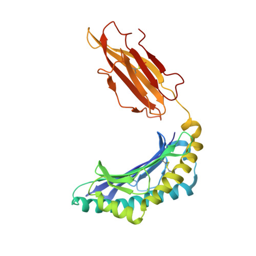
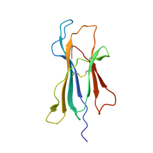
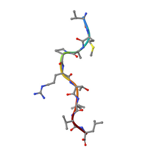
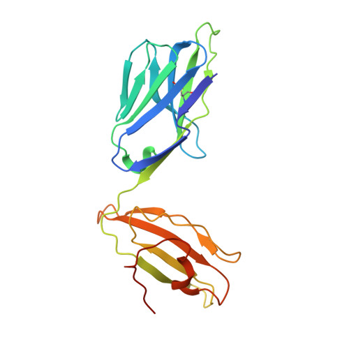
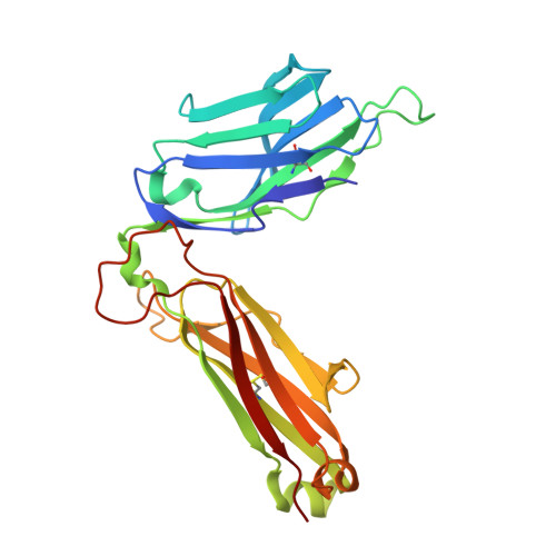
Structure of the HLA-E-VMAPRTLVL/GF4 TCR complex


Experimental Data Snapshot

  • Method: X-RAY DIFFRACTION
  • Resolution: 3.10 Å
  • R-Value Free: 
    0.263 (Depositor), 0.282 (DCC) 
  • R-Value Work: 
    0.215 (Depositor), 0.230 (DCC) 
  • R-Value Observed: 
    0.217 (Depositor) 

wwPDB Validation 3D Report Full Report

Validation slider image for 5W1W

This is version 1.3 of the entry. See complete history. 

Literature

A conserved energetic footprint underpins recognition of human leukocyte antigen-E by two distinct alpha beta T cell receptors.

Sullivan, L.C., Walpole, N.G., Farenc, C., Pietra, G., Sum, M.J.W., Clements, C.S., Lee, E.J., Beddoe, T., Falco, M., Mingari, M.C., Moretta, L., Gras, S., Rossjohn, J., Brooks, A.G.

(2017) J Biol Chem 292: 21149-21158

  • DOI: https://doi.org/10.1074/jbc.M117.807719
  • Primary Citation Related Structures: 
    5W1V, 5W1W

  • PubMed Abstract: 

    αβ T cell receptors (TCRs) interact with peptides bound to the polymorphic major histocompatibility complex class Ia (MHC-Ia) and class II (MHC-II) molecules as well as the essentially monomorphic MHC class Ib (MHC-Ib) molecules. Although there is a large amount of information on how TCRs engage with MHC-Ia and MHC-II, our understanding of TCR/MHC-Ib interactions is very limited. Infection with cytomegalovirus (CMV) can elicit a CD8 + T cell response restricted by the human MHC-Ib molecule human leukocyte antigen (HLA)-E and specific for an epitope from UL40 (VMAPRTLIL), which is characterized by biased TRBV14 gene usage. Here we describe an HLA-E-restricted CD8 + T cell able to recognize an allotypic variant of the UL40 peptide with a modification at position 8 (P8) of the peptide (VMAPRTLVL) that uses the TRBV9 gene segment. We report the structures of a TRBV9 + TCR in complex with the HLA-E molecule presenting the two peptides. Our data revealed that the TRBV9 + TCR adopts a different docking mode and molecular footprint atop HLA-E when compared with the TRBV14 + TCR-HLA-E ternary complex. Additionally, despite their differing V gene segment usage and different docking mechanisms, mutational analyses showed that the TCRs shared a conserved energetic footprint on the HLA-E molecule, focused around the peptide-binding groove. Hence, we provide new insights into how monomorphic MHC molecules interact with T cells.


  • Organizational Affiliation: 
    • From the Department of Microbiology and Immunology and Peter Doherty Institute for Infection and Immunity, University of Melbourne, Melbourne 3000, Australia.

Macromolecule Content 

  • Total Structure Weight: 381.04 kDa 
  • Atom Count: 26,478 
  • Modeled Residue Count: 3,300 
  • Deposited Residue Count: 3,360 
  • Unique protein chains: 5

Macromolecules

Find similar proteins by:|  3D Structure
Entity ID: 1
MoleculeChains  Sequence LengthOrganismDetailsImage
HLA class I histocompatibility antigen, alpha chain E
A, F, K, P
278Homo sapiensMutation(s): 0 
Gene Names: HLA-E, HLA-6.2, HLAE
UniProt & NIH Common Fund Data Resources
Find proteins for P13747 (Homo sapiens)
Explore P13747 
Go to UniProtKB:  P13747
PHAROS:  P13747
GTEx:  ENSG00000204592 
Entity Groups
Sequence Clusters30% Identity50% Identity70% Identity90% Identity95% Identity100% Identity
UniProt GroupP13747
Sequence Annotations
Expand
Reference Sequence
Find similar proteins by:|  3D Structure
Entity ID: 2
MoleculeChains  Sequence LengthOrganismDetailsImage
Beta-2-microglobulin
B, G, L, Q
100Homo sapiensMutation(s): 0 
Gene Names: B2M, CDABP0092, HDCMA22P
UniProt & NIH Common Fund Data Resources
Find proteins for P61769 (Homo sapiens)
Explore P61769 
Go to UniProtKB:  P61769
PHAROS:  P61769
GTEx:  ENSG00000166710 
Entity Groups
Sequence Clusters30% Identity50% Identity70% Identity90% Identity95% Identity100% Identity
UniProt GroupP61769
Sequence Annotations
Expand
Reference Sequence
Find similar proteins by:  Sequence   |   3D Structure  
Entity ID: 3
MoleculeChains  Sequence LengthOrganismDetailsImage
leader peptide of HLA class I histocompatibility antigen, A alpha chain
C, H, M, R
9Homo sapiensMutation(s): 0 
Gene Names: HLA-A
UniProt & NIH Common Fund Data Resources
Find proteins for P04439 (Homo sapiens)
Explore P04439 
Go to UniProtKB:  P04439
PHAROS:  P04439
GTEx:  ENSG00000206503 
Entity Groups
UniProt GroupP04439
Sequence Annotations
Expand
Reference Sequence
Find similar proteins by:|  3D Structure
Entity ID: 4
MoleculeChains  Sequence LengthOrganismDetailsImage
GF4 T cell receptor alpha chain
D, I, N, S
207Homo sapiensMutation(s): 0 
Gene Names: TRAC TCRA
Entity Groups
Sequence Clusters30% Identity50% Identity70% Identity90% Identity95% Identity100% Identity
Sequence Annotations
Expand
Reference Sequence
Find similar proteins by:|  3D Structure
Entity ID: 5
MoleculeChains  Sequence LengthOrganismDetailsImage
GF4 T cell receptor beta chain
E, J, O, T
246Homo sapiensMutation(s): 0 
Gene Names: TRBC1
Entity Groups
Sequence Clusters30% Identity50% Identity70% Identity90% Identity95% Identity100% Identity
Sequence Annotations
Expand
Reference Sequence

Experimental Data & Validation

Experimental Data

  • Method: X-RAY DIFFRACTION
  • Resolution: 3.10 Å
  • R-Value Free:  0.263 (Depositor), 0.282 (DCC) 
  • R-Value Work:  0.215 (Depositor), 0.230 (DCC) 
  • R-Value Observed: 0.217 (Depositor) 
Space Group: P 21 21 21
Unit Cell:
Length ( Å )Angle ( ˚ )
a = 73.371α = 90
b = 225.926β = 90
c = 276.267γ = 90
Software Package:
Software NamePurpose
BUSTERrefinement
XDSdata reduction
SCALAdata scaling

Structure Validation

View Full Validation Report


Entry History 

Deposition Data

Revision History  (Full details and data files)

  • Version 1.0: 2017-10-04
    Type: Initial release
  • Version 1.1: 2017-10-18
    Changes: Database references
  • Version 1.2: 2018-01-03
    Changes: Database references
  • Version 1.3: 2024-11-20
    Changes: Data collection, Database references, Structure summary