5VXY | pdb_00005vxy

Cryo-EM reconstruction of PAK pilus from Pseudomonas aeruginosa


Experimental Data Snapshot

  • Method: ELECTRON MICROSCOPY
  • Resolution: 8.00 Å
  • Aggregation State: FILAMENT 
  • Reconstruction Method: HELICAL 

wwPDB Validation 3D Report Full Report

Validation slider image for 5VXY

This is version 1.7 of the entry. See complete history

Literature

Cryoelectron Microscopy Reconstructions of the Pseudomonas aeruginosa and Neisseria gonorrhoeae Type IV Pili at Sub-nanometer Resolution.

Wang, F.Coureuil, M.Osinski, T.Orlova, A.Altindal, T.Gesbert, G.Nassif, X.Egelman, E.H.Craig, L.

(2017) Structure 25: 1423-1435.e4

  • DOI: https://doi.org/10.1016/j.str.2017.07.016
  • Primary Citation Related Structures: 
    5VXX, 5VXY

  • PubMed Abstract: 

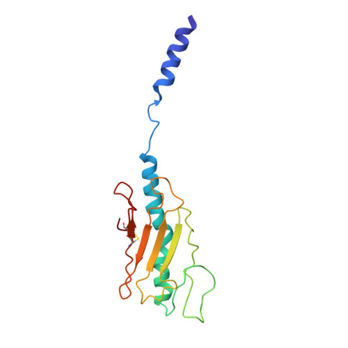
    We report here cryoelectron microscopy reconstructions of type IV pili (T4P) from two important human pathogens, Pseudomonas aeruginosa and Neisseria gonorrhoeae, at ∼ 8 and 5 Å resolution, respectively. The two structures reveal distinct arrangements of the pilin globular domains on the pilus surfaces, which impart different helical parameters, but similar packing of the conserved N-terminal α helices, α1, in the filament core. In contrast to the continuous α helix seen in the X-ray crystal structures of the P. aeruginosa and N. gonorrhoeae pilin subunits, α1 in the pilus filaments has a melted segment located between conserved helix-breaking residues Gly14 and Pro22, as seen for the Neisseria meningitidis T4P. Using mutagenesis we show that Pro22 is critical for pilus assembly, as are Thr2 and Glu5, which are positioned to interact in the hydrophobic filament core. These structures provide a framework for understanding T4P assembly, function, and biophysical properties.


  • Organizational Affiliation
    • Department of Biochemistry and Molecular Genetics, University of Virginia School of Medicine, Charlottesville, VA 22908, USA.

Macromolecule Content 

  • Total Structure Weight: 315.44 kDa 
  • Atom Count: 22,113 
  • Modeled Residue Count: 3,024 
  • Deposited Residue Count: 3,024 
  • Unique protein chains: 1

Macromolecules

Find similar proteins by:|  3D Structure
Entity ID: 1
MoleculeChains  Sequence LengthOrganismDetailsImage
Fimbrial protein
A, B, C, D, E
A, B, C, D, E, F, G, H, I, J, K, L, M, N, O, P, Q, R, S, T, U
144Pseudomonas aeruginosa PAKMutation(s): 0 
Membrane Entity: Yes 
UniProt
Find proteins for P02973 (Pseudomonas aeruginosa)
Explore P02973 
Go to UniProtKB:  P02973
Entity Groups
Sequence Clusters30% Identity50% Identity70% Identity90% Identity95% Identity100% Identity
UniProt GroupP02973
Sequence Annotations
Expand
Reference Sequence

Experimental Data & Validation

Experimental Data

  • Method: ELECTRON MICROSCOPY
  • Resolution: 8.00 Å
  • Aggregation State: FILAMENT 
  • Reconstruction Method: HELICAL 
EM Software:
TaskSoftware PackageVersion
RECONSTRUCTIONSPIDER
MODEL REFINEMENTPHENIX
MODEL REFINEMENTCoot

Structure Validation

View Full Validation Report


Entry History 

& Funding Information

Deposition Data


Funding OrganizationLocationGrant Number
National Institutes of Health/National Institute Of Allergy and Infectious Diseases (NIH/NIAID)United StatesAI114902
Canadian Institutes of Health Research (CIHR)CanadaMOP125959
French National Research AgencyFranceANR14CE14001002
Fondation pour la Recherche MedicaleFrance--

Revision History  (Full details and data files)

  • Version 1.0: 2017-07-12
    Type: Initial release
  • Version 1.1: 2017-09-13
    Changes: Author supporting evidence, Data collection
  • Version 1.2: 2017-09-20
    Changes: Database references
  • Version 1.3: 2017-10-04
    Changes: Author supporting evidence
  • Version 1.4: 2018-07-18
    Changes: Data collection
  • Version 1.5: 2019-10-02
    Changes: Author supporting evidence, Data collection, Derived calculations, Experimental preparation
  • Version 1.6: 2019-12-11
    Changes: Author supporting evidence
  • Version 1.7: 2024-11-20
    Changes: Data collection, Database references, Structure summary