5VVT

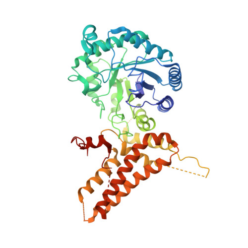
Structural Investigations of the Substrate Specificity of Human O-GlcNAcase


Experimental Data Snapshot

  • Method: X-RAY DIFFRACTION
  • Resolution: 2.80 Å
  • R-Value Free: 0.291 
  • R-Value Work: 0.211 
  • R-Value Observed: 0.215 

Starting Model: experimental
View more details

wwPDB Validation   3D Report Full Report


Ligand Structure Quality Assessment 


This is version 1.3 of the entry. See complete history


Literature

Structural insights into the substrate binding adaptability and specificity of human O-GlcNAcase.

Li, B.Li, H.Hu, C.W.Jiang, J.

(2017) Nat Commun 8: 666-666

  • DOI: https://doi.org/10.1038/s41467-017-00865-1
  • Primary Citation of Related Structures:  
    5VVO, 5VVT, 5VVU, 5VVV, 5VVX

  • PubMed Abstract: 

    The O-linked β-N-acetyl glucosamine (O-GlcNAc) modification dynamically regulates the functions of numerous proteins. A single human enzyme O-linked β-N-acetyl glucosaminase (O-GlcNAcase or OGA) hydrolyzes this modification. To date, it remains largely unknown how OGA recognizes various substrates. Here we report the structures of OGA in complex with each of four distinct glycopeptide substrates that contain a single O-GlcNAc modification on a serine or threonine residue. Intriguingly, these glycopeptides bind in a bidirectional yet conserved conformation within the substrate-binding cleft of OGA. This study provides fundamental insights into a general principle that confers the substrate binding adaptability and specificity to OGA in O-GlcNAc regulation.O-linked β-N-acetyl glucosamine (O-GlcNAc) is an important protein modification that is hydrolyzed by O-GlcNAcase (OGA). Here the authors give insights into OGA substrate recognition by presenting four human OGA structures complexed with glycopeptide substrates containing a single O-GlcNAc modification on either a serine or threonine.


  • Organizational Affiliation

    Pharmaceutical Sciences Division, School of Pharmacy, University of Wisconsin-Madison, Madison, WI, 53705, USA.


Macromolecules
Find similar proteins by:  (by identity cutoff)  |  3D Structure
Entity ID: 1
MoleculeChains Sequence LengthOrganismDetailsImage
Protein O-GlcNAcase
A, C
504Homo sapiensMutation(s): 1 
Gene Names: MGEA5HEXCKIAA0679MEA5
EC: 3.2.1.169 (PDB Primary Data), 3.2.1 (PDB Primary Data)
UniProt & NIH Common Fund Data Resources
Find proteins for O60502 (Homo sapiens)
Explore O60502 
Go to UniProtKB:  O60502
PHAROS:  O60502
GTEx:  ENSG00000198408 
Entity Groups  
Sequence Clusters30% Identity50% Identity70% Identity90% Identity95% Identity100% Identity
UniProt GroupO60502
Sequence Annotations
Expand
  • Reference Sequence

Find similar proteins by:  Sequence   |   3D Structure  

Entity ID: 2
MoleculeChains Sequence LengthOrganismDetailsImage
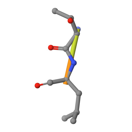
ELK1 peptide
B, D
8Homo sapiensMutation(s): 0 
UniProt & NIH Common Fund Data Resources
Find proteins for P19419 (Homo sapiens)
Explore P19419 
Go to UniProtKB:  P19419
PHAROS:  P19419
GTEx:  ENSG00000126767 
Entity Groups  
UniProt GroupP19419
Glycosylation
Glycosylation Sites: 1Go to GlyGen: P19419-1
Sequence Annotations
Expand
  • Reference Sequence
Small Molecules
Ligands 1 Unique
IDChains Name / Formula / InChI Key2D Diagram3D Interactions
NAG
Query on NAG

Download Ideal Coordinates CCD File 
E [auth B],
F [auth D]
2-acetamido-2-deoxy-beta-D-glucopyranose
C8 H15 N O6
OVRNDRQMDRJTHS-FMDGEEDCSA-N
Experimental Data & Validation

Experimental Data

  • Method: X-RAY DIFFRACTION
  • Resolution: 2.80 Å
  • R-Value Free: 0.291 
  • R-Value Work: 0.211 
  • R-Value Observed: 0.215 
  • Space Group: P 1 21 1
Unit Cell:
Length ( Å )Angle ( ˚ )
a = 82.363α = 90
b = 95.793β = 114.51
c = 89.323γ = 90
Software Package:
Software NamePurpose
REFMACrefinement
HKL-2000data reduction
HKL-2000data scaling
PHASERphasing

Structure Validation

View Full Validation Report


Ligand Structure Quality Assessment 


Entry History & Funding Information

Deposition Data


Funding OrganizationLocationGrant Number
University of Wisconsin-MadisonUnited States--

Revision History  (Full details and data files)

  • Version 1.0: 2017-09-27
    Type: Initial release
  • Version 1.1: 2017-10-04
    Changes: Database references
  • Version 1.2: 2020-07-29
    Type: Remediation
    Reason: Carbohydrate remediation
    Changes: Data collection, Derived calculations, Structure summary
  • Version 1.3: 2023-10-04
    Changes: Data collection, Database references, Refinement description, Structure summary