Structural and functional characterization of FabG4 from Mycolicibacterium smegmatis.
Ran, X., Parikh, P., Abendroth, J., Arakaki, T.L., Clifton, M.C., Edwards, T.E., Lorimer, D.D., Mayclin, S., Staker, B.L., Myler, P., McLaughlin, K.J.(2024) Acta Crystallogr F Struct Biol Commun 80: 82-91
- PubMed: 38656226 Search on PubMedSearch on PubMed Central
- DOI: https://doi.org/10.1107/S2053230X2400356X
- Primary Citation Related Structures:
3U0B, 5VP5 - PubMed Abstract:
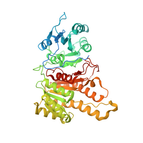
The rise in antimicrobial resistance is a global health crisis and necessitates the development of novel strategies to treat infections. For example, in 2022 tuberculosis (TB) was the second leading infectious killer after COVID-19, with multi-drug-resistant strains of TB having an ∼40% fatality rate. Targeting essential biosynthetic pathways in pathogens has proven to be successful for the development of novel antimicrobial treatments. Fatty-acid synthesis (FAS) in bacteria proceeds via the type II pathway, which is substantially different from the type I pathway utilized in animals. This makes bacterial fatty-acid biosynthesis (Fab) enzymes appealing as drug targets. FabG is an essential FASII enzyme, and some bacteria, such as Mycobacterium tuberculosis, the causative agent of TB, harbor multiple homologs. FabG4 is a conserved, high-molecular-weight FabG (HMwFabG) that was first identified in M. tuberculosis and is distinct from the canonical low-molecular-weight FabG. Here, structural and functional analyses of Mycolicibacterium smegmatis FabG4, the third HMwFabG studied to date, are reported. Crystal structures of NAD + and apo MsFabG4, along with kinetic analyses, show that MsFabG4 preferentially binds and uses NADH when reducing CoA substrates. As M. smegmatis is often used as a model organism for M. tuberculosis, these studies may aid the development of drugs to treat TB and add to the growing body of research that distinguish HMwFabGs from the archetypal low-molecular-weight FabG.
- Department of Chemistry, Vassar College, 124 Raymond Avenue, Poughkeepsie, NY 12604, USA.
Organizational Affiliation: