5VNY | pdb_00005vny

Crystal structure of DM14-3 domain of Lgd


Experimental Data Snapshot

  • Method: X-RAY DIFFRACTION
  • Resolution: 1.10 Å
  • R-Value Free: 
    0.178 (Depositor), 0.179 (DCC) 
  • R-Value Work: 
    0.170 (Depositor), 0.171 (DCC) 
  • R-Value Observed: 
    0.170 (Depositor) 

wwPDB Validation 3D Report Full Report

Validation slider image for 5VNY

This is version 1.1 of the entry. See complete history

Literature

Structural Basis for Regulation of ESCRT-III Complexes by Lgd.

McMillan, B.J.Tibbe, C.Drabek, A.A.Seegar, T.C.M.Blacklow, S.C.Klein, T.

(2017) Cell Rep 19: 1750-1757

  • DOI: https://doi.org/10.1016/j.celrep.2017.05.026
  • Primary Citation Related Structures: 
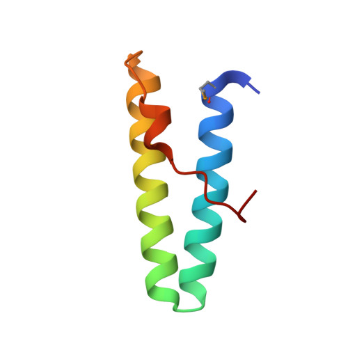
    5VNY, 5VO5

  • PubMed Abstract: 

    The ESCRT-III complex induces outward membrane budding and fission through homotypic polymerization of its core component Shrub/CHMP4B. Shrub activity is regulated by its direct interaction with a protein called Lgd in flies, or CC2D1A or B in humans. Here, we report the structural basis for this interaction and propose a mechanism for regulation of polymer assembly. The isolated third DM14 repeat of Lgd binds Shrub, and an Lgd fragment containing only this DM14 repeat and its C-terminal C2 domain is sufficient for in vivo function. The DM14 domain forms a helical hairpin with a conserved, positively charged tip, that, in the structure of a DM14 domain-Shrub complex, occupies a negatively charged surface of Shrub that is otherwise used for homopolymerization. Lgd mutations at this interface disrupt its function in flies, confirming functional importance. Together, these data argue that Lgd regulates ESCRT activity by controlling access to the Shrub self-assembly surface.


  • Organizational Affiliation
    • Department of Biological Chemistry and Molecular Pharmacology, Harvard Medical School, Boston, MA 02115, USA.

Macromolecule Content 

  • Total Structure Weight: 7.43 kDa 
  • Atom Count: 632 
  • Modeled Residue Count: 65 
  • Deposited Residue Count: 66 
  • Unique protein chains: 1

Macromolecules

Find similar proteins by:|  3D Structure
Entity ID: 1
MoleculeChains  Sequence LengthOrganismDetailsImage
Lethal (2) giant discs 1, isoform B66Drosophila melanogasterMutation(s): 0 
Gene Names: l(2)gd1CG4713Dmel_CG4713
UniProt
Find proteins for Q9VKJ9 (Drosophila melanogaster)
Explore Q9VKJ9 
Go to UniProtKB:  Q9VKJ9
Entity Groups
Sequence Clusters30% Identity50% Identity70% Identity90% Identity95% Identity100% Identity
UniProt GroupQ9VKJ9
Sequence Annotations
Expand
Reference Sequence

Small Molecules

Modified Residues  1 Unique
IDChains TypeFormula2D DiagramParent
MSE
Query on MSE
A
L-PEPTIDE LINKINGC5 H11 N O2 SeMET

Experimental Data & Validation

Experimental Data

  • Method: X-RAY DIFFRACTION
  • Resolution: 1.10 Å
  • R-Value Free:  0.178 (Depositor), 0.179 (DCC) 
  • R-Value Work:  0.170 (Depositor), 0.171 (DCC) 
  • R-Value Observed: 0.170 (Depositor) 
Space Group: P 41 21 2
Unit Cell:
Length ( Å )Angle ( ˚ )
a = 29.67α = 90
b = 29.67β = 90
c = 139.11γ = 90
Software Package:
Software NamePurpose
PHENIXrefinement
XDSdata reduction
XDSdata scaling
PHENIXphasing

Structure Validation

View Full Validation Report


Entry History 

Deposition Data

Revision History  (Full details and data files)

  • Version 1.0: 2017-06-14
    Type: Initial release
  • Version 1.1: 2024-11-20
    Changes: Data collection, Database references, Structure summary