5VNP | pdb_00005vnp

X-ray crystal structure of Halotag bound to the P1 benzoxadiazole fluorogenic ligand


Experimental Data Snapshot

  • Method: X-RAY DIFFRACTION
  • Resolution: 2.23 Å
  • R-Value Free: 
    0.229 (Depositor), 0.233 (DCC) 
  • R-Value Work: 
    0.198 (Depositor), 0.205 (DCC) 
  • R-Value Observed: 
    0.199 (Depositor) 

Starting Model: experimental
View more details

wwPDB Validation 3D Report Full Report

Validation slider image for 5VNP

Ligand Structure Quality Assessment 


This is version 1.4 of the entry. See complete history

Literature

AgHalo: A Facile Fluorogenic Sensor to Detect Drug-Induced Proteome Stress.

Liu, Y.Fares, M.Dunham, N.P.Gao, Z.Miao, K.Jiang, X.Bollinger, S.S.Boal, A.K.Zhang, X.

(2017) Angew Chem Int Ed Engl 56: 8672-8676

  • DOI: https://doi.org/10.1002/anie.201702417
  • Primary Citation Related Structures: 
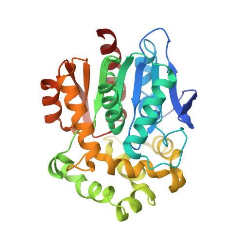
    5VNP

  • PubMed Abstract: 

    Drug-induced proteome stress that involves protein aggregation may cause adverse effects and undermine the safety profile of a drug. Safety of drugs is regularly evaluated using cytotoxicity assays that measure cell death. However, these assays provide limited insights into the presence of proteome stress in live cells. A fluorogenic protein sensor is reported to detect drug-induced proteome stress prior to cell death. An aggregation prone Halo-tag mutant (AgHalo) was evolved to sense proteome stress through its aggregation. Detection of such conformational changes was enabled by a fluorogenic ligand that fluoresces upon AgHalo forming soluble aggregates. Using 5 common anticancer drugs, we exemplified detection of differential proteome stress before any cell death was observed. Thus, this sensor can be used to evaluate drug safety in a regime that the current cytotoxicity assays cannot cover and be generally applied to detect proteome stress induced by other toxins.


  • Organizational Affiliation
    • Department of Chemistry, The Pennsylvania State University, University Park, PA, 16802, USA.

Macromolecule Content 

  • Total Structure Weight: 66.87 kDa 
  • Atom Count: 4,803 
  • Modeled Residue Count: 580 
  • Deposited Residue Count: 580 
  • Unique protein chains: 1

Macromolecules

Find similar proteins by:|  3D Structure
Entity ID: 1
MoleculeChains  Sequence LengthOrganismDetailsImage
Haloalkane dehalogenase
A, B
290Rhodococcus rhodochrousMutation(s): 0 
Gene Names: dhaA
EC: 3.8.1.5
UniProt
Find proteins for P0A3G2 (Rhodococcus rhodochrous)
Explore P0A3G2 
Go to UniProtKB:  P0A3G2
Entity Groups
Sequence Clusters30% Identity50% Identity70% Identity90% Identity95% Identity100% Identity
UniProt GroupP0A3G2
Sequence Annotations
Expand
Reference Sequence

Experimental Data & Validation

Experimental Data

  • Method: X-RAY DIFFRACTION
  • Resolution: 2.23 Å
  • R-Value Free:  0.229 (Depositor), 0.233 (DCC) 
  • R-Value Work:  0.198 (Depositor), 0.205 (DCC) 
  • R-Value Observed: 0.199 (Depositor) 
Space Group: P 1 21 1
Unit Cell:
Length ( Å )Angle ( ˚ )
a = 44.272α = 90
b = 69.481β = 95.82
c = 88.95γ = 90
Software Package:
Software NamePurpose
HKL-2000data scaling
REFMACrefinement
PDB_EXTRACTdata extraction
HKL-2000data reduction
PHASERphasing
DENZOdata reduction
SCALEPACKdata scaling

Structure Validation

View Full Validation Report


Ligand Structure Quality Assessment 


Entry History 

Deposition Data

Revision History  (Full details and data files)

  • Version 1.0: 2017-06-14
    Type: Initial release
  • Version 1.1: 2017-07-26
    Changes: Database references
  • Version 1.2: 2017-11-22
    Changes: Refinement description
  • Version 1.3: 2023-10-04
    Changes: Data collection, Database references, Refinement description
  • Version 1.4: 2024-10-23
    Changes: Structure summary