5VN0 | pdb_00005vn0

Water-forming NADH oxidase from Lactobacillus brevis (LbNOX) bound to NADH.


Experimental Data Snapshot

  • Method: X-RAY DIFFRACTION
  • Resolution: 2.00 Å
  • R-Value Free: 
    0.225 (Depositor), 0.226 (DCC) 
  • R-Value Work: 
    0.179 (Depositor), 0.182 (DCC) 
  • R-Value Observed: 
    0.182 (Depositor) 

Starting Model: experimental
View more details

wwPDB Validation 3D Report Full Report

Validation slider image for 5VN0

Ligand Structure Quality Assessment 


This is version 1.4 of the entry. See complete history

Literature

A genetically encoded tool for manipulation of NADP(+)/NADPH in living cells.

Cracan, V.Titov, D.V.Shen, H.Grabarek, Z.Mootha, V.K.

(2017) Nat Chem Biol 13: 1088-1095

  • DOI: https://doi.org/10.1038/nchembio.2454
  • Primary Citation Related Structures: 
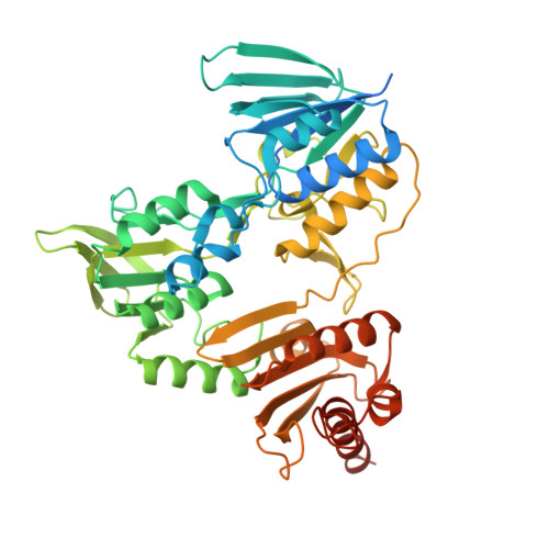
    5VN0, 5VOH

  • PubMed Abstract: 

    The redox coenzymes NADH and NADPH are broadly required for energy metabolism, biosynthesis and detoxification. Despite detailed knowledge of specific enzymes and pathways that utilize these coenzymes, a holistic understanding of the regulation and compartmentalization of NADH- and NADPH-dependent pathways is lacking, partly because of a lack of tools with which to investigate these processes in living cells. We have previously reported the use of the naturally occurring Lactobacillus brevis H 2 O-forming NADH oxidase (LbNOX) as a genetic tool for manipulation of the NAD + /NADH ratio in human cells. Here, we present triphosphopyridine nucleotide oxidase (TPNOX), a rationally designed and engineered mutant of LbNOX that is strictly specific to NADPH. We characterized the effects of TPNOX expression on cellular metabolism and used it in combination with LbNOX to show how the redox states of mitochondrial NADPH and NADH pools are connected.


  • Organizational Affiliation
    • Howard Hughes Medical Institute and Department of Molecular Biology, Massachusetts General Hospital, Boston, Massachusetts, USA.

Macromolecule Content 

  • Total Structure Weight: 461.7 kDa 
  • Atom Count: 30,845 
  • Modeled Residue Count: 3,593 
  • Deposited Residue Count: 4,144 
  • Unique protein chains: 1

Macromolecules

Find similar proteins by:|  3D Structure
Entity ID: 1
MoleculeChains  Sequence LengthOrganismDetailsImage
NAD(FAD)-dependent dehydrogenase
A, B, C, D, E
A, B, C, D, E, F, G, H
518Levilactobacillus brevis ATCC 367Mutation(s): 0 
Gene Names: LVIS_1558
UniProt
Find proteins for Q03Q85 (Levilactobacillus brevis (strain ATCC 367 / BCRC 12310 / CIP 105137 / JCM 1170 / LMG 11437 / NCIMB 947 / NCTC 947))
Explore Q03Q85 
Go to UniProtKB:  Q03Q85
Entity Groups
Sequence Clusters30% Identity50% Identity70% Identity90% Identity95% Identity100% Identity
UniProt GroupQ03Q85
Sequence Annotations
Expand
Reference Sequence

Small Molecules

Ligands 4 Unique
IDChains Name / Formula / InChI Key2D Diagram3D Interactions
FAD

Query on FAD


Download:Ideal Coordinates CCD File
AA [auth E]
EA [auth F]
I [auth A]
KA [auth G]
N [auth B]
AA [auth E],
EA [auth F],
I [auth A],
KA [auth G],
N [auth B],
OA [auth H],
R [auth C],
V [auth D]
FLAVIN-ADENINE DINUCLEOTIDE
C27 H33 N9 O15 P2
VWWQXMAJTJZDQX-UYBVJOGSSA-N
NAI

Query on NAI


Download:Ideal Coordinates CCD File
CA [auth E]
GA [auth F]
K [auth A]
MA [auth G]
P [auth B]
CA [auth E],
GA [auth F],
K [auth A],
MA [auth G],
P [auth B],
QA [auth H],
T [auth C],
X [auth D]
1,4-DIHYDRONICOTINAMIDE ADENINE DINUCLEOTIDE
C21 H29 N7 O14 P2
BOPGDPNILDQYTO-NNYOXOHSSA-N
EDO

Query on EDO


Download:Ideal Coordinates CCD File
DA [auth E]
HA [auth F]
IA [auth F]
JA [auth F]
L [auth A]
DA [auth E],
HA [auth F],
IA [auth F],
JA [auth F],
L [auth A],
M [auth A],
NA [auth G],
Q [auth B],
RA [auth H],
SA [auth H],
U [auth C],
Y [auth D],
Z [auth D]
1,2-ETHANEDIOL
C2 H6 O2
LYCAIKOWRPUZTN-UHFFFAOYSA-N
OXY

Query on OXY


Download:Ideal Coordinates CCD File
BA [auth E]
FA [auth F]
J [auth A]
LA [auth G]
O [auth B]
BA [auth E],
FA [auth F],
J [auth A],
LA [auth G],
O [auth B],
PA [auth H],
S [auth C],
W [auth D]
OXYGEN MOLECULE
O2
MYMOFIZGZYHOMD-UHFFFAOYSA-N

Experimental Data & Validation

Experimental Data

  • Method: X-RAY DIFFRACTION
  • Resolution: 2.00 Å
  • R-Value Free:  0.225 (Depositor), 0.226 (DCC) 
  • R-Value Work:  0.179 (Depositor), 0.182 (DCC) 
  • R-Value Observed: 0.182 (Depositor) 
Space Group: P 1
Unit Cell:
Length ( Å )Angle ( ˚ )
a = 96.525α = 63.37
b = 107.511β = 90.04
c = 119.298γ = 89.95
Software Package:
Software NamePurpose
PHENIXrefinement
HKL-2000data scaling
HKL-2000data reduction
PHASERphasing
PDB_EXTRACTdata extraction

Structure Validation

View Full Validation Report


Ligand Structure Quality Assessment 


Entry History 

& Funding Information

Deposition Data


Funding OrganizationLocationGrant Number
National Institutes of Health/National Institute of General Medical Sciences (NIH/NIGMS)United StatesR01GM099683

Revision History  (Full details and data files)

  • Version 1.0: 2017-08-02
    Type: Initial release
  • Version 1.1: 2017-08-30
    Changes: Database references, Refinement description
  • Version 1.2: 2017-09-27
    Changes: Database references
  • Version 1.3: 2020-01-01
    Changes: Author supporting evidence
  • Version 1.4: 2023-10-04
    Changes: Data collection, Database references, Refinement description