5VMG

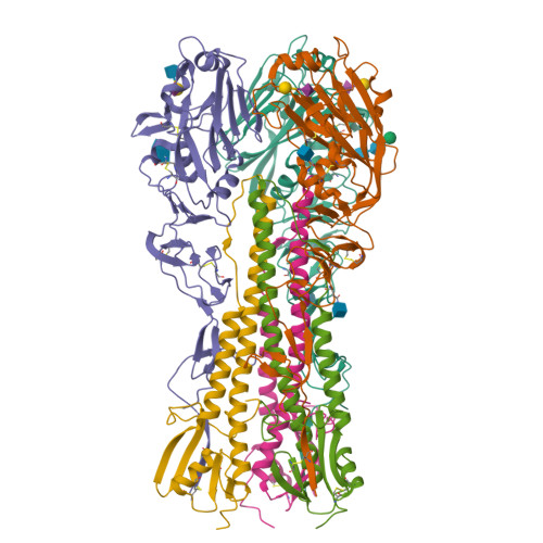
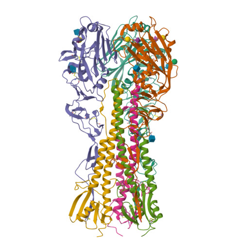
Influenza hemagglutinin H1 mutant DH1E in complex with 6'SLN


Experimental Data Snapshot

  • Method: X-RAY DIFFRACTION
  • Resolution: 2.45 Å
  • R-Value Free: 0.225 
  • R-Value Work: 0.191 
  • R-Value Observed: 0.193 

Starting Model: experimental
View more details

wwPDB Validation   3D Report Full Report


Ligand Structure Quality Assessment 


This is version 2.3 of the entry. See complete history


Literature

Determinant of receptor-preference switch in influenza hemagglutinin.

Ni, F.Kondrashkina, E.Wang, Q.

(2017) Virology 513: 98-107

  • DOI: https://doi.org/10.1016/j.virol.2017.10.010
  • Primary Citation of Related Structures:  
    5VMC, 5VMF, 5VMG, 5VMJ

  • PubMed Abstract: 

    Influenza pandemic occurs when a new strain from other animal species overcomes the inter-species barriers and supports rapid human-to-human transmission. A critical prerequisite to this process is that hemagglutinin (HA) acquires a few key mutations to switch from avian receptors to human receptors. Previous studies suggest that H1 and H2/H3 HAs use different sets of mutations for the switch. This report shows that HA from the 1918 H1N1 pandemic virus (1918H1 HA) adopts the set of mutations used by H2/H3 HAs in receptor-preference switch when its 130-loop is made similar to those of H2/H3 HAs. Thus, the 130-loop appears to be the key determinant for the different mutations employed by pandemic H1 or H2/H3 HA. The correlation of the mutational routes and the 130-loop as unraveled in this study opens the door for efficient investigation of mutations required by other HA subtypes for inter-human airborne transmission.


  • Organizational Affiliation

    Verna and Marrs McLean Department of Biochemistry and Molecular Biology, Baylor College of Medicine, One Baylor Plaza, Houston, TX 77030, USA.


Macromolecules
Find similar proteins by:  (by identity cutoff)  |  3D Structure
Entity ID: 1
MoleculeChains Sequence LengthOrganismDetailsImage
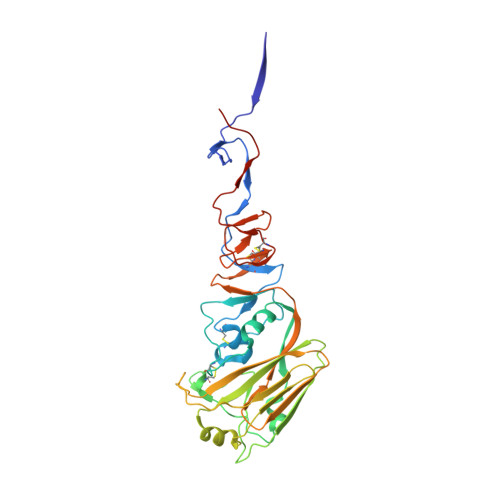
Hemagglutinin HA1
A, C, E
326Influenza A virus (A/New_York/1/18(H1N1))Mutation(s): 3 
Gene Names: HA
UniProt
Find proteins for Q9WFX4 (Influenza A virus)
Explore Q9WFX4 
Go to UniProtKB:  Q9WFX4
Entity Groups  
Sequence Clusters30% Identity50% Identity70% Identity90% Identity95% Identity100% Identity
UniProt GroupQ9WFX4
Glycosylation
Glycosylation Sites: 2
Sequence Annotations
Expand
  • Reference Sequence
Find similar proteins by:  (by identity cutoff)  |  3D Structure
Entity ID: 2
MoleculeChains Sequence LengthOrganismDetailsImage
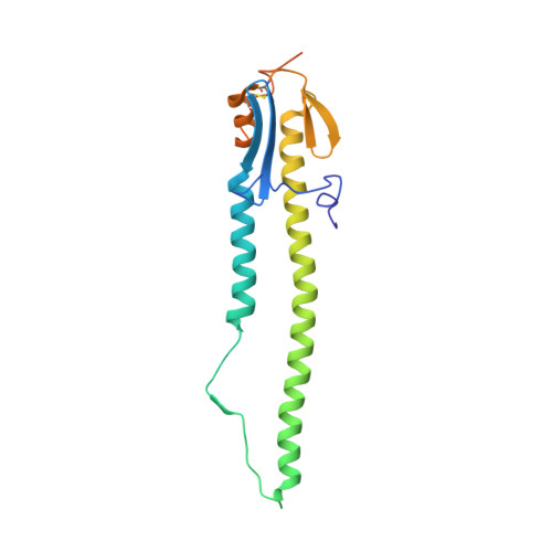
Hemagglutinin HA2
B, D, F
191Influenza A virus (A/Brevig Mission/1/1918(H1N1))Mutation(s): 0 
Gene Names: HA
UniProt
Find proteins for Q9WFX3 (Influenza A virus (strain A/Brevig Mission/1/1918 H1N1))
Explore Q9WFX3 
Go to UniProtKB:  Q9WFX3
Entity Groups  
Sequence Clusters30% Identity50% Identity70% Identity90% Identity95% Identity100% Identity
UniProt GroupQ9WFX3
Sequence Annotations
Expand
  • Reference Sequence
Oligosaccharides

Help

Entity ID: 3
MoleculeChains Length2D Diagram Glycosylation3D Interactions
beta-D-mannopyranose-(1-4)-2-acetamido-2-deoxy-beta-D-glucopyranose-(1-4)-2-acetamido-2-deoxy-beta-D-glucopyranose
G
3N-Glycosylation
Glycosylation Resources
GlyTouCan:  G15407YE
GlyCosmos:  G15407YE
GlyGen:  G15407YE
Entity ID: 4
MoleculeChains Length2D Diagram Glycosylation3D Interactions
N-acetyl-alpha-neuraminic acid-(2-6)-beta-D-galactopyranose
H, I
2N/A
Glycosylation Resources
GlyTouCan:  G63069TR
GlyCosmos:  G63069TR
GlyGen:  G63069TR
Entity ID: 5
MoleculeChains Length2D Diagram Glycosylation3D Interactions
2-acetamido-2-deoxy-beta-D-glucopyranose-(1-4)-2-acetamido-2-deoxy-beta-D-glucopyranose
J
2N-Glycosylation
Glycosylation Resources
GlyTouCan:  G42666HT
GlyCosmos:  G42666HT
GlyGen:  G42666HT
Entity ID: 6
MoleculeChains Length2D Diagram Glycosylation3D Interactions
N-acetyl-alpha-neuraminic acid-(2-6)-beta-D-galactopyranose-(1-4)-2-acetamido-2-deoxy-beta-D-glucopyranose
K
3N/A
Glycosylation Resources
GlyTouCan:  G73578JC
GlyCosmos:  G73578JC
GlyGen:  G73578JC
Biologically Interesting Molecules (External Reference) 1 Unique
Experimental Data & Validation

Experimental Data

  • Method: X-RAY DIFFRACTION
  • Resolution: 2.45 Å
  • R-Value Free: 0.225 
  • R-Value Work: 0.191 
  • R-Value Observed: 0.193 
  • Space Group: P 1 21 1
Unit Cell:
Length ( Å )Angle ( ˚ )
a = 95.38α = 90
b = 81.37β = 95.45
c = 120.96γ = 90
Software Package:
Software NamePurpose
PHENIXrefinement
MOSFLMdata reduction
SCALAdata scaling
PHENIXphasing

Structure Validation

View Full Validation Report


Ligand Structure Quality Assessment 


Entry History & Funding Information

Deposition Data


Funding OrganizationLocationGrant Number
National Institutes of Health/National Institute Of Allergy and Infectious Diseases (NIH/NIAID)United StatesR01-AI067839
National Institutes of Health/National Institute of General Medical Sciences (NIH/NIGMS)United StatesR01-GM116280

Revision History  (Full details and data files)

  • Version 1.0: 2017-11-08
    Type: Initial release
  • Version 2.0: 2020-07-29
    Type: Remediation
    Reason: Carbohydrate remediation
    Changes: Advisory, Atomic model, Data collection, Derived calculations, Structure summary
  • Version 2.1: 2022-03-23
    Changes: Author supporting evidence, Database references, Derived calculations, Structure summary
  • Version 2.2: 2023-10-04
    Changes: Data collection, Refinement description
  • Version 2.3: 2024-10-16
    Changes: Structure summary