


Crystal structure of adenylosuccinate lyase ADE13 from Candida albicans
Stogios, P.J.To be published.
Experimental Data Snapshot
Starting Model: experimental
View more details
wwPDB Validation 3D Report Full Report

Entity ID: 1 | |||||
|---|---|---|---|---|---|
| Molecule | Chains | Sequence Length | Organism | Details | Image |
| Adenylosuccinate lyase | 482 | Candida albicans SC5314 | Mutation(s): 0 Gene Names: ADE13, CAALFM_CR06150CA, orf19.3870 EC: 4.3.2.2 | 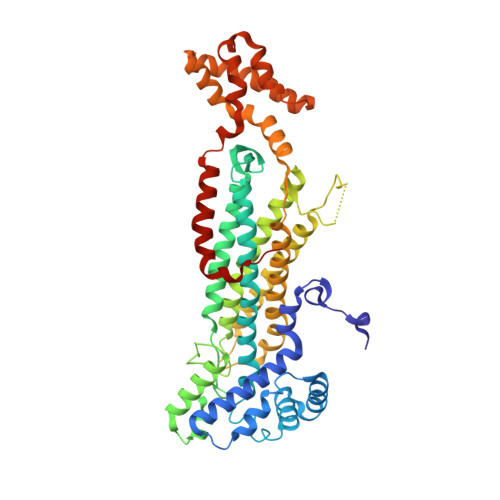 | |
UniProt | |||||
Find proteins for A0A1D8PT56 (Candida albicans (strain SC5314 / ATCC MYA-2876)) Explore A0A1D8PT56 Go to UniProtKB: A0A1D8PT56 | |||||
Entity Groups | |||||
| Sequence Clusters | 30% Identity50% Identity70% Identity90% Identity95% Identity100% Identity | ||||
| UniProt Group | A0A1D8PT56 | ||||
Sequence AnnotationsExpand | |||||
Reference Sequence | |||||
| Ligands 5 Unique | |||||
|---|---|---|---|---|---|
| ID | Chains | Name / Formula / InChI Key | 2D Diagram | 3D Interactions | |
| PGE Download:Ideal Coordinates CCD File | P [auth B] | TRIETHYLENE GLYCOL C6 H14 O4 ZIBGPFATKBEMQZ-UHFFFAOYSA-N |  | ||
| GOL Download:Ideal Coordinates CCD File | F [auth A], M [auth B] | GLYCEROL C3 H8 O3 PEDCQBHIVMGVHV-UHFFFAOYSA-N |  | ||
| CA Download:Ideal Coordinates CCD File | N [auth B], O [auth B] | CALCIUM ION Ca BHPQYMZQTOCNFJ-UHFFFAOYSA-N |  | ||
| CL Download:Ideal Coordinates CCD File | C [auth A] D [auth A] E [auth A] H [auth B] I [auth B] | CHLORIDE ION Cl VEXZGXHMUGYJMC-UHFFFAOYSA-M |  | ||
| MG Download:Ideal Coordinates CCD File | G [auth A], Q [auth B] | MAGNESIUM ION Mg JLVVSXFLKOJNIY-UHFFFAOYSA-N |  | ||
| Length ( Å ) | Angle ( ˚ ) |
|---|---|
| a = 85.03 | α = 90 |
| b = 157.845 | β = 90 |
| c = 374.554 | γ = 90 |
| Software Name | Purpose |
|---|---|
| PHENIX | refinement |
| HKL-3000 | data reduction |
| HKL-3000 | data scaling |
| PHENIX | phasing |
| PHENIX | model building |
| Coot | model building |
| Funding Organization | Location | Grant Number |
|---|---|---|
| National Institutes of Health/National Institute Of Allergy and Infectious Diseases (NIH/NIAID) | United States | HHSCN27220120026C |


