5VKK

Crystal structure of Fab fragment of anti-CD22 Epratuzumab


Experimental Data Snapshot

  • Method: X-RAY DIFFRACTION
  • Resolution: 2.01 Å
  • R-Value Free: 0.237 
  • R-Value Work: 0.212 
  • R-Value Observed: 0.213 

Starting Model: experimental
View more details

wwPDB Validation   3D Report Full Report


This is version 1.5 of the entry. See complete history


Literature

Molecular basis of human CD22 function and therapeutic targeting.

Ereno-Orbea, J.Sicard, T.Cui, H.Mazhab-Jafari, M.T.Benlekbir, S.Guarne, A.Rubinstein, J.L.Julien, J.P.

(2017) Nat Commun 8: 764-764

  • DOI: https://doi.org/10.1038/s41467-017-00836-6
  • Primary Citation of Related Structures:  
    5VKJ, 5VKK, 5VKM, 5VL3

  • PubMed Abstract: 

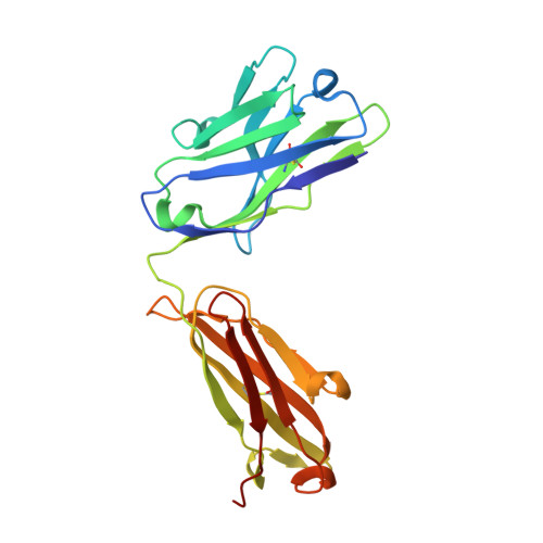
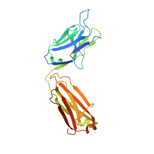
    CD22 maintains a baseline level of B-cell inhibition to keep humoral immunity in check. As a B-cell-restricted antigen, CD22 is targeted in therapies against dysregulated B cells that cause autoimmune diseases and blood cancers. Here we report the crystal structure of human CD22 at 2.1 Å resolution, which reveals that specificity for α2-6 sialic acid ligands is dictated by a pre-formed β-hairpin as a unique mode of recognition across sialic acid-binding immunoglobulin-type lectins. The CD22 ectodomain adopts an extended conformation that facilitates concomitant CD22 nanocluster formation on B cells and binding to trans ligands to avert autoimmunity in mammals. We structurally delineate the CD22 site targeted by the therapeutic antibody epratuzumab at 3.1 Å resolution and determine a critical role for CD22 N-linked glycosylation in antibody engagement. Our studies provide molecular insights into mechanisms governing B-cell inhibition and valuable clues for the design of immune modulators in B-cell dysfunction.The B-cell-specific co-receptor CD22 is a therapeutic target for depleting dysregulated B cells. Here the authors structurally characterize the ectodomain of CD22 and present its crystal structure with the bound therapeutic antibody epratuzumab, which gives insights into the mechanism of inhibition of B-cell activation.


  • Organizational Affiliation

    Program in Molecular Medicine, The Hospital for Sick Children Research Institute, Toronto, ON, Canada, M5G 0A4.


Macromolecules
Find similar proteins by:  (by identity cutoff)  |  3D Structure
Entity ID: 1
MoleculeChains Sequence LengthOrganismDetailsImage
Epratuzumab Fab Heavy ChainA [auth H],
C [auth A]
219Mus musculusHomo sapiens
This entity is chimeric
Mutation(s): 0 
UniProt
Find proteins for Q6N089 (Homo sapiens)
Explore Q6N089 
Go to UniProtKB:  Q6N089
Entity Groups  
Sequence Clusters30% Identity50% Identity70% Identity90% Identity95% Identity100% Identity
UniProt GroupQ6N089
Sequence Annotations
Expand
  • Reference Sequence
Find similar proteins by:  (by identity cutoff)  |  3D Structure
Entity ID: 2
MoleculeChains Sequence LengthOrganismDetailsImage
Epratuzumab Fab Light ChainB [auth L],
D [auth B]
219Mus musculusHomo sapiens
This entity is chimeric
Mutation(s): 0 
UniProt
Find proteins for Q8TCD0 (Homo sapiens)
Explore Q8TCD0 
Go to UniProtKB:  Q8TCD0
Entity Groups  
Sequence Clusters30% Identity50% Identity70% Identity90% Identity95% Identity100% Identity
UniProt GroupQ8TCD0
Sequence Annotations
Expand
  • Reference Sequence
Experimental Data & Validation

Experimental Data

  • Method: X-RAY DIFFRACTION
  • Resolution: 2.01 Å
  • R-Value Free: 0.237 
  • R-Value Work: 0.212 
  • R-Value Observed: 0.213 
  • Space Group: P 1
Unit Cell:
Length ( Å )Angle ( ˚ )
a = 56.712α = 71.81
b = 61.564β = 81.07
c = 65.285γ = 75.95
Software Package:
Software NamePurpose
PHENIXrefinement
XSCALEdata scaling
PDB_EXTRACTdata extraction
XDSdata reduction
PHASERphasing

Structure Validation

View Full Validation Report


Entry History & Funding Information

Deposition Data


Funding OrganizationLocationGrant Number
Canadian Institutes of Health Research (CIHR)Canada111111
Canadian Institutes of Health Research (CIHR)CanadaPJT-148811
Canadian Institutes of Health Research (CIHR)CanadaBPF-144483

Revision History  (Full details and data files)

  • Version 1.0: 2017-10-04
    Type: Initial release
  • Version 1.1: 2017-10-11
    Changes: Database references, Source and taxonomy
  • Version 1.2: 2017-10-25
    Changes: Database references
  • Version 1.3: 2018-01-31
    Changes: Structure summary
  • Version 1.4: 2020-01-08
    Changes: Author supporting evidence
  • Version 1.5: 2023-10-04
    Changes: Data collection, Database references, Refinement description