5VJ1 | pdb_00005vj1

Crystal structure of a Pseudomonas malonate decarboxylase hetero-tetramer in complex with coenzyme A


Experimental Data Snapshot

  • Method: X-RAY DIFFRACTION
  • Resolution: 3.00 Å
  • R-Value Free: 
    0.233 (Depositor), 0.206 (DCC) 
  • R-Value Work: 
    0.173 (Depositor) 
  • R-Value Observed: 
    0.176 (Depositor) 

wwPDB Validation 3D Report Full Report

Validation slider image for 5VJ1

Ligand Structure Quality Assessment 


This is version 1.1 of the entry. See complete history

Literature

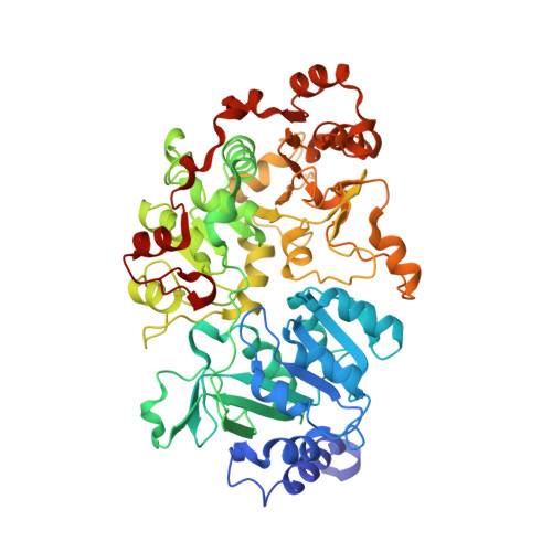
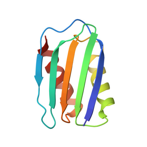
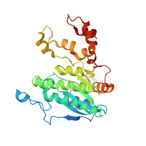
Crystal structure of a Pseudomonas malonate decarboxylase holoenzyme hetero-tetramer.

Maderbocus, R.Fields, B.L.Hamilton, K.Luo, S.Tran, T.H.Dietrich, L.E.P.Tong, L.

(2017) Nat Commun 8: 160-160

  • DOI: https://doi.org/10.1038/s41467-017-00233-z
  • Primary Citation Related Structures: 
    5VIP, 5VIT, 5VJ1

  • PubMed Abstract: 

    Pseudomonas species and other aerobic bacteria have a biotin-independent malonate decarboxylase that is crucial for their utilization of malonate as the sole carbon and energy source. The malonate decarboxylase holoenzyme contains four subunits, having an acyl-carrier protein (MdcC subunit) with a distinct prosthetic group, as well as decarboxylase (MdcD-MdcE) and acyl-carrier protein transferase (MdcA) catalytic activities. Here we report the crystal structure of a Pseudomonas malonate decarboxylase hetero-tetramer, as well as biochemical and functional studies based on the structural information. We observe a malonate molecule in the active site of MdcA and we also determine the structure of malonate decarboxylase with CoA in the active site of MdcD-MdcE. Both structures provide molecular insights into malonate decarboxylase catalysis. Mutations in the hetero-tetramer interface can abolish holoenzyme formation. Mutations in the hetero-tetramer interface and the active sites can abolish Pseudomonas aeruginosa growth in a defined medium with malonate as the sole carbon source.Some aerobic bacteria contain a biotin-independent malonate decarboxylase (MDC), which allows them to use malonate as the sole carbon source. Here, the authors present the crystal structure of a Pseudomonas MDC and give insights into its catalytic mechanism and function.


  • Organizational Affiliation
    • Department of Biological Sciences, Columbia University, New York, NY, 10027, USA.

Macromolecule Content 

  • Total Structure Weight: 267.4 kDa 
  • Atom Count: 18,197 
  • Modeled Residue Count: 2,368 
  • Deposited Residue Count: 2,448 
  • Unique protein chains: 4

Macromolecules

Find similar proteins by:|  3D Structure
Entity ID: 1
MoleculeChains  Sequence LengthOrganismDetailsImage
MdcAA,
E [auth I]
554Pseudomonas aeruginosa PAO1Mutation(s): 0 
Gene Names: mdcAPA0208
UniProt
Find proteins for Q9I6T0 (Pseudomonas aeruginosa (strain ATCC 15692 / DSM 22644 / CIP 104116 / JCM 14847 / LMG 12228 / 1C / PRS 101 / PAO1))
Explore Q9I6T0 
Go to UniProtKB:  Q9I6T0
Entity Groups
Sequence Clusters30% Identity50% Identity70% Identity90% Identity95% Identity100% Identity
UniProt GroupQ9I6T0
Sequence Annotations
Expand
Reference Sequence
Find similar proteins by:|  3D Structure
Entity ID: 2
MoleculeChains  Sequence LengthOrganismDetailsImage
MdcCB [auth C],
F [auth K]
99Pseudomonas protegens Pf-5Mutation(s): 0 
Gene Names: mdcCPFL_5818
UniProt
Find proteins for Q4K4F7 (Pseudomonas fluorescens (strain ATCC BAA-477 / NRRL B-23932 / Pf-5))
Explore Q4K4F7 
Go to UniProtKB:  Q4K4F7
Entity Groups
Sequence Clusters30% Identity50% Identity70% Identity90% Identity95% Identity100% Identity
UniProt GroupQ4K4F7
Sequence Annotations
Expand
Reference Sequence
Find similar proteins by:|  3D Structure
Entity ID: 3
MoleculeChains  Sequence LengthOrganismDetailsImage
MdcDC [auth D],
G [auth L]
287Pseudomonas aeruginosaMutation(s): 0 
Gene Names: madCmdcDAO964_31600AOY09_06294BH593_13640PAERUG_E15_London_28_01_14_07061PAERUG_P32_London_17_VIM_2_10_11_04127PAMH19_0209
EC: 2.1.3.10 (PDB Primary Data), 2.1.3.1 (PDB Primary Data)
UniProt
Find proteins for Q9I6S7 (Pseudomonas aeruginosa (strain ATCC 15692 / DSM 22644 / CIP 104116 / JCM 14847 / LMG 12228 / 1C / PRS 101 / PAO1))
Explore Q9I6S7 
Go to UniProtKB:  Q9I6S7
Entity Groups
Sequence Clusters30% Identity50% Identity70% Identity90% Identity95% Identity100% Identity
UniProt GroupQ9I6S7
Sequence Annotations
Expand
Reference Sequence
Find similar proteins by:|  3D Structure
Entity ID: 4
MoleculeChains  Sequence LengthOrganismDetailsImage
MdcED [auth E],
H [auth M]
284Pseudomonas aeruginosaMutation(s): 0 
Gene Names: madDAO964_31595AOY09_06293PAERUG_E15_London_28_01_14_07062PAERUG_P32_London_17_VIM_2_10_11_04128
EC: 2.1.3.10
UniProt
Find proteins for Q9I6S6 (Pseudomonas aeruginosa (strain ATCC 15692 / DSM 22644 / CIP 104116 / JCM 14847 / LMG 12228 / 1C / PRS 101 / PAO1))
Explore Q9I6S6 
Go to UniProtKB:  Q9I6S6
Entity Groups
Sequence Clusters30% Identity50% Identity70% Identity90% Identity95% Identity100% Identity
UniProt GroupQ9I6S6
Sequence Annotations
Expand
Reference Sequence

Experimental Data & Validation

Experimental Data

  • Method: X-RAY DIFFRACTION
  • Resolution: 3.00 Å
  • R-Value Free:  0.233 (Depositor), 0.206 (DCC) 
  • R-Value Work:  0.173 (Depositor) 
  • R-Value Observed: 0.176 (Depositor) 
Space Group: P 1 21 1
Unit Cell:
Length ( Å )Angle ( ˚ )
a = 98.74α = 90
b = 163.56β = 94.01
c = 100.44γ = 90
Software Package:
Software NamePurpose
PHENIXrefinement
XDSdata reduction
XDSdata scaling

Structure Validation

View Full Validation Report


Ligand Structure Quality Assessment 


Entry History 

Deposition Data

Revision History  (Full details and data files)

  • Version 1.0: 2017-08-16
    Type: Initial release
  • Version 1.1: 2024-03-13
    Changes: Data collection, Database references