5VGJ | pdb_00005vgj

Crystal Structure of the Human Fab VRC38.01, an HIV-1 V1V2-Directed Neutralizing Antibody Isolated from Donor N90, bound to a scaffolded WITO V1V2 domain


Experimental Data Snapshot

  • Method: X-RAY DIFFRACTION
  • Resolution: 3.46 Å
  • R-Value Free: 
    0.273 (Depositor), 0.275 (DCC) 
  • R-Value Work: 
    0.214 (Depositor), 0.214 (DCC) 
  • R-Value Observed: 
    0.217 (Depositor) 

Starting Model: experimental
View more details

wwPDB Validation 3D Report Full Report

Validation slider image for 5VGJ

Ligand Structure Quality Assessment 


This is version 2.2 of the entry. See complete history

Literature

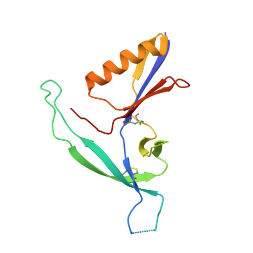
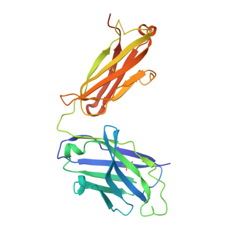
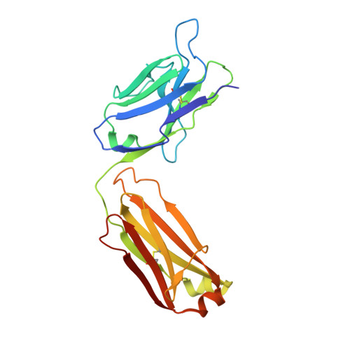
Virus-like Particles Identify an HIV V1V2 Apex-Binding Neutralizing Antibody that Lacks a Protruding Loop.

Cale, E.M.Gorman, J.Radakovich, N.A.Crooks, E.T.Osawa, K.Tong, T.Li, J.Nagarajan, R.Ozorowski, G.Ambrozak, D.R.Asokan, M.Bailer, R.T.Bennici, A.K.Chen, X.Doria-Rose, N.A.Druz, A.Feng, Y.Joyce, M.G.Louder, M.K.O'Dell, S.Oliver, C.Pancera, M.Connors, M.Hope, T.J.Kepler, T.B.Wyatt, R.T.Ward, A.B.Georgiev, I.S.Kwong, P.D.Mascola, J.R.Binley, J.M.

(2017) Immunity 46: 777-791.e10

  • DOI: https://doi.org/10.1016/j.immuni.2017.04.011
  • Primary Citation Related Structures: 
    5EWI, 5VGJ

  • PubMed Abstract: 

    Most HIV-1-specific neutralizing antibodies isolated to date exhibit unusual characteristics that complicate their elicitation. Neutralizing antibodies that target the V1V2 apex of the HIV-1 envelope (Env) trimer feature unusually long protruding loops, which enable them to penetrate the HIV-1 glycan shield. As antibodies with loops of requisite length are created through uncommon recombination events, an alternative mode of apex binding has been sought. Here, we isolated a lineage of Env apex-directed neutralizing antibodies, N90-VRC38.01-11, by using virus-like particles and conformationally stabilized Env trimers as B cell probes. A crystal structure of N90-VRC38.01 with a scaffolded V1V2 revealed a binding mode involving side-chain-to-side-chain interactions that reduced the distance the antibody loop must traverse the glycan shield, thereby facilitating V1V2 binding via a non-protruding loop. The N90-VRC38 lineage thus identifies a solution for V1V2-apex binding that provides a more conventional B cell pathway for vaccine design.


  • Organizational Affiliation
    • Vaccine Research Center, National Institute of Allergy and Infectious Diseases, National Institutes of Health, Bethesda, MD 20892, USA.

Macromolecule Content 

  • Total Structure Weight: 67.64 kDa 
  • Atom Count: 4,520 
  • Modeled Residue Count: 560 
  • Deposited Residue Count: 590 
  • Unique protein chains: 3

Macromolecules

Find similar proteins by:|  3D Structure
Entity ID: 1
MoleculeChains  Sequence LengthOrganismDetailsImage
1FD6-V1V2-WITOA [auth G]134Human immunodeficiency virus 1Mutation(s): 0 
UniProt
Find proteins for Q5G5U5 (Human immunodeficiency virus type 1)
Explore Q5G5U5 
Go to UniProtKB:  Q5G5U5
Entity Groups
Sequence Clusters30% Identity50% Identity70% Identity90% Identity95% Identity100% Identity
UniProt GroupQ5G5U5
Glycosylation
Glycosylation Sites: 4
Sequence Annotations
Expand
Reference Sequence
Find similar proteins by:|  3D Structure
Entity ID: 2
MoleculeChains  Sequence LengthOrganismDetailsImage
VRC38.01 Fab Heavy ChainB [auth H]236Homo sapiensMutation(s): 0 
Find similar proteins by:|  3D Structure
Entity ID: 3
MoleculeChains  Sequence LengthOrganismDetailsImage
VRC38.01 Fab Light ChainC [auth L]220Homo sapiensMutation(s): 0 

Oligosaccharides

Help  
Entity ID: 4
MoleculeChains Length2D Diagram GlycosylationD Interactions
alpha-D-mannopyranose-(1-3)-[alpha-D-mannopyranose-(1-6)]alpha-D-mannopyranose-(1-6)-[alpha-D-mannopyranose-(1-3)]beta-D-mannopyranose-(1-4)-2-acetamido-2-deoxy-beta-D-glucopyranose-(1-4)-2-acetamido-2-deoxy-beta-D-glucopyranoseD [auth A],
E [auth B]
7N-Glycosylation
Glycosylation Resources
GlyTouCan: G55220VL
GlyCosmos: G55220VL
GlyGen: G55220VL
Entity ID: 5
MoleculeChains Length2D Diagram GlycosylationD Interactions
alpha-D-mannopyranose-(1-3)-[alpha-D-mannopyranose-(1-6)]beta-D-mannopyranose-(1-4)-2-acetamido-2-deoxy-beta-D-glucopyranose-(1-4)-2-acetamido-2-deoxy-beta-D-glucopyranoseF [auth C]5N-Glycosylation
Glycosylation Resources
GlyTouCan: G22768VO
GlyCosmos: G22768VO
GlyGen: G22768VO

Small Molecules

Ligands 1 Unique
IDChains Name / Formula / InChI Key2D Diagram3D Interactions
NAG

Query on NAG


Download:Ideal Coordinates CCD File
G
2-acetamido-2-deoxy-beta-D-glucopyranose
C8 H15 N O6
OVRNDRQMDRJTHS-FMDGEEDCSA-N

Experimental Data & Validation

Experimental Data

  • Method: X-RAY DIFFRACTION
  • Resolution: 3.46 Å
  • R-Value Free:  0.273 (Depositor), 0.275 (DCC) 
  • R-Value Work:  0.214 (Depositor), 0.214 (DCC) 
  • R-Value Observed: 0.217 (Depositor) 
Space Group: P 32 2 1
Unit Cell:
Length ( Å )Angle ( ˚ )
a = 106.572α = 90
b = 106.572β = 90
c = 129.857γ = 120
Software Package:
Software NamePurpose
PHENIXrefinement
HKL-2000data scaling
PDB_EXTRACTdata extraction
HKL-2000data reduction
PHENIXphasing
DENZOdata reduction
SCALEPACKdata scaling

Structure Validation

View Full Validation Report


Ligand Structure Quality Assessment 


Entry History 

Deposition Data

Revision History  (Full details and data files)

  • Version 1.0: 2017-05-31
    Type: Initial release
  • Version 1.1: 2017-11-22
    Changes: Refinement description
  • Version 2.0: 2020-07-29
    Type: Remediation
    Reason: Carbohydrate remediation
    Changes: Advisory, Atomic model, Data collection, Derived calculations, Structure summary
  • Version 2.1: 2023-10-04
    Changes: Data collection, Database references, Derived calculations, Refinement description, Structure summary
  • Version 2.2: 2024-11-20
    Changes: Structure summary