


Crystal structure of D-isomer specific 2-hydroxyacid dehydrogenase from Xanthobacter autotrophicus Py2 in complex with NADPH and MES.
Lipowska, J., Shabalin, I.G., Almo, S.C., Minor, W.To be published.
Experimental Data Snapshot
Starting Model: experimental
View more details
Entity ID: 1 | |||||
|---|---|---|---|---|---|
| Molecule | Chains | Sequence Length | Organism | Details | Image |
| D-isomer specific 2-hydroxyacid dehydrogenase NAD-binding | 341 | Xanthobacter versatilis | Mutation(s): 0 Gene Names: Xaut_2571 | 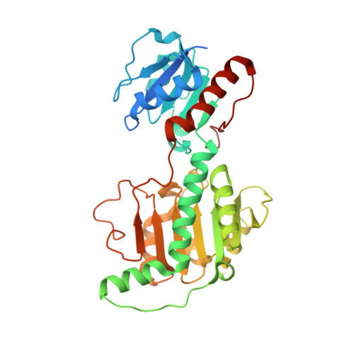 | |
UniProt | |||||
Entity Groups | |||||
| Sequence Clusters | 30% Identity50% Identity70% Identity90% Identity95% Identity100% Identity | ||||
| UniProt Group | A7IIH0 | ||||
Sequence AnnotationsExpand | |||||
Reference Sequence | |||||
| Ligands 2 Unique | |||||
|---|---|---|---|---|---|
| ID | Chains | Name / Formula / InChI Key | 2D Diagram | 3D Interactions | |
| NDP Download:Ideal Coordinates CCD File | BA [auth J] K [auth A] N [auth B] P [auth C] R [auth D] | NADPH DIHYDRO-NICOTINAMIDE-ADENINE-DINUCLEOTIDE PHOSPHATE C21 H30 N7 O17 P3 ACFIXJIJDZMPPO-NNYOXOHSSA-N |  | ||
| MES Download:Ideal Coordinates CCD File | AA [auth I] CA [auth J] L [auth A] M [auth A] O [auth B] | 2-(N-MORPHOLINO)-ETHANESULFONIC ACID C6 H13 N O4 S SXGZJKUKBWWHRA-UHFFFAOYSA-N |  | ||
| Length ( Å ) | Angle ( ˚ ) |
|---|---|
| a = 132.514 | α = 90 |
| b = 311.036 | β = 90 |
| c = 98.977 | γ = 90 |
| Software Name | Purpose |
|---|---|
| HKL-3000 | data collection |
| HKL-3000 | data reduction |
| HKL-3000 | data scaling |
| HKL-3000 | phasing |
| MOLREP | phasing |
| REFMAC | refinement |
| PDB_EXTRACT | data extraction |
| SCALEPACK | data scaling |
| Funding Organization | Location | Grant Number |
|---|---|---|
| National Institutes of Health/National Institute of General Medical Sciences (NIH/NIGMS) | United States | -- |


