5VF6 | pdb_00005vf6

Crystal structure of single chain variable fragment (scFv45).


Experimental Data Snapshot

  • Method: X-RAY DIFFRACTION
  • Resolution: 1.64 Å
  • R-Value Free: 
    0.196 (Depositor), 0.199 (DCC) 
  • R-Value Work: 
    0.172 (Depositor), 0.173 (DCC) 
  • R-Value Observed: 
    0.173 (Depositor) 

Starting Model: experimental
View more details

wwPDB Validation 3D Report Full Report

Validation slider image for 5VF6

This is version 1.3 of the entry. See complete history

Literature

Harnessing insulin- and leptin-induced oxidation of PTP1B for therapeutic development.

Krishnan, N.Bonham, C.A.Rus, I.A.Shrestha, O.K.Gauss, C.M.Haque, A.Tocilj, A.Joshua-Tor, L.Tonks, N.K.

(2018) Nat Commun 9: 283-283

  • DOI: https://doi.org/10.1038/s41467-017-02252-2
  • Primary Citation Related Structures: 
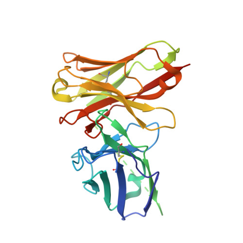
    5VF6

  • PubMed Abstract: 

    The protein tyrosine phosphatase PTP1B is a major regulator of glucose homeostasis and energy metabolism, and a validated target for therapeutic intervention in diabetes and obesity. Nevertheless, it is a challenging target for inhibitor development. Previously, we generated a recombinant antibody (scFv45) that recognizes selectively the oxidized, inactive conformation of PTP1B. Here, we provide a molecular basis for its interaction with reversibly oxidized PTP1B. Furthermore, we have identified a small molecule inhibitor that mimics the effects of scFv45. Our data provide proof-of-concept that stabilization of PTP1B in an inactive, oxidized conformation by small molecules can promote insulin and leptin signaling. This work illustrates a novel paradigm for inhibiting the signaling function of PTP1B that may be exploited for therapeutic intervention in diabetes and obesity.


  • Organizational Affiliation
    • Cold Spring Harbor Laboratory, 1 Bungtown Road, Cold Spring Harbor, NY, 11724, USA.

Macromolecule Content 

  • Total Structure Weight: 25.56 kDa 
  • Atom Count: 1,993 
  • Modeled Residue Count: 226 
  • Deposited Residue Count: 250 
  • Unique protein chains: 1

Macromolecules

Find similar proteins by:|  3D Structure
Entity ID: 1
MoleculeChains  Sequence LengthOrganismDetailsImage
single chain variable fragment250Gallus gallusMutation(s): 0 

Experimental Data & Validation

Experimental Data

  • Method: X-RAY DIFFRACTION
  • Resolution: 1.64 Å
  • R-Value Free:  0.196 (Depositor), 0.199 (DCC) 
  • R-Value Work:  0.172 (Depositor), 0.173 (DCC) 
  • R-Value Observed: 0.173 (Depositor) 
Space Group: P 32 2 1
Unit Cell:
Length ( Å )Angle ( ˚ )
a = 97.232α = 90
b = 97.232β = 90
c = 61.26γ = 120
Software Package:
Software NamePurpose
PHENIXrefinement
XDSdata reduction
XDSdata scaling
PHASERphasing

Structure Validation

View Full Validation Report


Entry History 

& Funding Information

Deposition Data


Funding OrganizationLocationGrant Number
National Institutes of Health/National Institute of General Medical Sciences (NIH/NIGMS)United StatesGM55989

Revision History  (Full details and data files)

  • Version 1.0: 2018-01-31
    Type: Initial release
  • Version 1.1: 2020-01-01
    Changes: Author supporting evidence
  • Version 1.2: 2023-10-04
    Changes: Data collection, Database references, Refinement description
  • Version 1.3: 2024-10-09
    Changes: Structure summary