5VEG | pdb_00005veg

Structure of a Short-Chain Flavodoxin Associated with a Non-Canonical PDU Bacterial Microcompartment


Experimental Data Snapshot

  • Method: X-RAY DIFFRACTION
  • Resolution: 1.99 Å
  • R-Value Free: 
    0.250 (Depositor), 0.251 (DCC) 
  • R-Value Work: 
    0.201 (Depositor), 0.204 (DCC) 
  • R-Value Observed: 
    0.204 (Depositor) 

Starting Model: experimental
View more details

wwPDB Validation 3D Report Full Report

Validation slider image for 5VEG

Ligand Structure Quality Assessment 


This is version 1.3 of the entry. See complete history

Literature

Structural and Functional Characterization of a Short-Chain Flavodoxin Associated with a Noncanonical 1,2-Propanediol Utilization Bacterial Microcompartment.

Plegaria, J.S.Sutter, M.Ferlez, B.Aussignargues, C.Niklas, J.Poluektov, O.G.Fromwiller, C.TerAvest, M.Utschig, L.M.Tiede, D.M.Kerfeld, C.A.

(2017) Biochemistry 56: 5679-5690

  • DOI: https://doi.org/10.1021/acs.biochem.7b00682
  • Primary Citation Related Structures: 
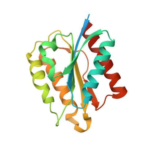
    5VEG

  • PubMed Abstract: 

    Bacterial microcompartments (BMCs) are proteinaceous organelles that encapsulate enzymes involved in CO 2 fixation (carboxysomes) or carbon catabolism (metabolosomes). Metabolosomes share a common core of enzymes and a distinct signature enzyme for substrate degradation that defines the function of the BMC (e.g., propanediol or ethanolamine utilization BMCs, or glycyl-radical enzyme microcompartments). Loci encoding metabolosomes also typically contain genes for proteins that support organelle function, such as regulation, transport of substrate, and cofactor (e.g., vitamin B 12 ) synthesis and recycling. Flavoproteins are frequently among these ancillary gene products, suggesting that these redox active proteins play an undetermined function in many metabolosomes. Here, we report the first characterization of a BMC-associated flavodoxin (Fld1C), a small flavoprotein, derived from the noncanonical 1,2-propanediol utilization BMC locus (PDU1C) of Lactobacillus reuteri. The 2.0 Å X-ray structure of Fld1C displays the α/β flavodoxin fold, which noncovalently binds a single flavin mononucleotide molecule. Fld1C is a short-chain flavodoxin with redox potentials of -240 ± 3 mV oxidized/semiquinone and -344 ± 1 mV semiquinone/hydroquinone versus the standard hydrogen electrode at pH 7.5. It can participate in an electron transfer reaction with a photoreductant to form a stable semiquinone species. Collectively, our structural and functional results suggest that PDU1C BMCs encapsulate Fld1C to store and transfer electrons for the reactivation and/or recycling of the B 12 cofactor utilized by the signature enzyme.


  • Organizational Affiliation
    • MSU-DOE Plant Research Laboratory, Michigan State University , East Lansing, Michigan 48824, United States.

Macromolecule Content 

  • Total Structure Weight: 59.79 kDa 
  • Atom Count: 3,702 
  • Modeled Residue Count: 445 
  • Deposited Residue Count: 534 
  • Unique protein chains: 1

Macromolecules

Find similar proteins by:|  3D Structure
Entity ID: 1
MoleculeChains  Sequence LengthOrganismDetailsImage
Flavodoxin
A, B, C
178Limosilactobacillus reuteriMutation(s): 0 
Gene Names: fldAA9P83_08720
UniProt
Find proteins for A5VM92 (Limosilactobacillus reuteri (strain DSM 20016))
Explore A5VM92 
Go to UniProtKB:  A5VM92
Entity Groups
Sequence Clusters30% Identity50% Identity70% Identity90% Identity95% Identity100% Identity
UniProt GroupA5VM92
Sequence Annotations
Expand
Reference Sequence

Small Molecules

Ligands 3 Unique
IDChains Name / Formula / InChI Key2D Diagram3D Interactions
FMN

Query on FMN


Download:Ideal Coordinates CCD File
D [auth A],
G [auth B],
K [auth C]
FLAVIN MONONUCLEOTIDE
C17 H21 N4 O9 P
FVTCRASFADXXNN-SCRDCRAPSA-N
CD

Query on CD


Download:Ideal Coordinates CCD File
E [auth A],
H [auth B],
I [auth B]
CADMIUM ION
Cd
WLZRMCYVCSSEQC-UHFFFAOYSA-N
NA

Query on NA


Download:Ideal Coordinates CCD File
F [auth A]
J [auth B]
L [auth C]
M [auth C]
N [auth C]
F [auth A],
J [auth B],
L [auth C],
M [auth C],
N [auth C],
O [auth C]
SODIUM ION
Na
FKNQFGJONOIPTF-UHFFFAOYSA-N

Experimental Data & Validation

Experimental Data

  • Method: X-RAY DIFFRACTION
  • Resolution: 1.99 Å
  • R-Value Free:  0.250 (Depositor), 0.251 (DCC) 
  • R-Value Work:  0.201 (Depositor), 0.204 (DCC) 
  • R-Value Observed: 0.204 (Depositor) 
Space Group: P 21 21 21
Unit Cell:
Length ( Å )Angle ( ˚ )
a = 47.301α = 90
b = 95.807β = 90
c = 102.378γ = 90
Software Package:
Software NamePurpose
PHENIXrefinement
XDSdata reduction
SCALAdata scaling
PHASERphasing

Structure Validation

View Full Validation Report


Ligand Structure Quality Assessment 


Entry History 

Deposition Data

Revision History  (Full details and data files)

  • Version 1.0: 2017-10-11
    Type: Initial release
  • Version 1.1: 2017-10-18
    Changes: Database references
  • Version 1.2: 2017-11-08
    Changes: Database references
  • Version 1.3: 2023-10-04
    Changes: Data collection, Database references, Derived calculations, Refinement description