5VC8 | pdb_00005vc8

Crystal structure of the WHSC1 PWWP1 domain


Experimental Data Snapshot

  • Method: X-RAY DIFFRACTION
  • Resolution: 1.80 Å
  • R-Value Free: 
    0.229 (Depositor), 0.234 (DCC) 
  • R-Value Work: 
    0.200 (Depositor), 0.209 (DCC) 
  • R-Value Observed: 
    0.201 (Depositor) 

wwPDB Validation 3D Report Full Report

Validation slider image for 5VC8

This is version 1.2 of the entry. See complete history

Literature

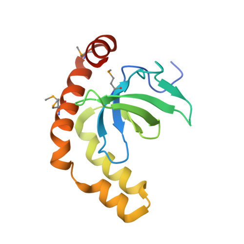
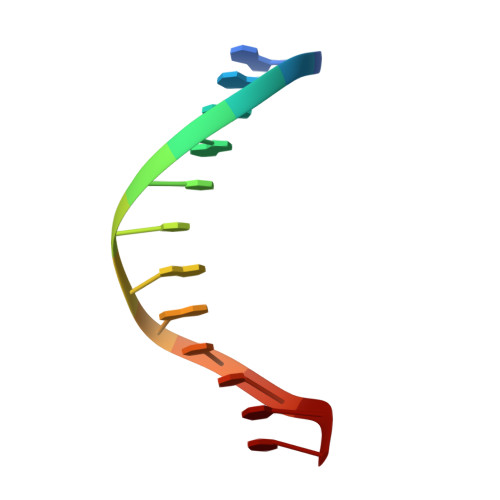
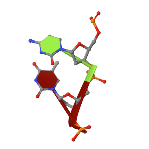
Histone and DNA binding ability studies of the NSD subfamily of PWWP domains.

Zhang, M.Yang, Y.Zhou, M.Dong, A.Yan, X.Loppnau, P.Min, J.Liu, Y.

(2021) Biochem Biophys Res Commun 569: 199-206

  • DOI: https://doi.org/10.1016/j.bbrc.2021.07.017
  • Primary Citation Related Structures: 
    4RXJ, 5VC8

  • PubMed Abstract: 

    The NSD proteins, namely NSD1, NSD2 and NSD3, are lysine methyltransferases, which catalyze mono- and di-methylation of histone H3K36. They are multi-domain proteins, including two PWWP domains (PWWP1 and PWWP2) separated by some other domains. These proteins act as potent oncoproteins and are implicated in various cancers. However the biological functions of these PWWP domains are still largely unknown. To better understand the functions of these proteins' PWWP domains, we cloned, expressed and purified all the PWWP domains of these NSD proteins to characterize their interactions with methylated histone peptides and dsDNA by quantitative binding assays and crystallographic analysis. Our studies indicate that all these PWWP domains except NSD1_PWWP1 bind to trimethylated H3K36, H3K79 peptides and dsDNA weakly. Our crystal structures uncover that the NDS3_PWWP2 and NSD2_PWWP1 domains, which hold an extremely long α-helix and α-helix bundle, respectively, need a conformation adjustment to interact with nucleosome.


  • Organizational Affiliation
    • College of Pharmaceutical Sciences, Soochow University, Suzhou, Jiangsu, China.

Macromolecule Content 

  • Total Structure Weight: 44.13 kDa 
  • Atom Count: 2,860 
  • Modeled Residue Count: 291 
  • Deposited Residue Count: 321 
  • Unique protein chains: 1
  • Unique nucleic acid chains: 2

Macromolecules


Find similar proteins by:|  3D Structure
Entity ID: 1
MoleculeChains  Sequence LengthOrganismDetailsImage
Histone-lysine N-methyltransferase NSD2
A, B
141Homo sapiensMutation(s): 0 
Gene Names: NSD2KIAA1090MMSETTRX5WHSC1
EC: 2.1.1.43 (PDB Primary Data), 2.1.1.357 (UniProt)
UniProt & NIH Common Fund Data Resources
Find proteins for O96028 (Homo sapiens)
Explore O96028 
Go to UniProtKB:  O96028
PHAROS:  O96028
GTEx:  ENSG00000109685 
Entity Groups
Sequence Clusters30% Identity50% Identity70% Identity90% Identity95% Identity100% Identity
UniProt GroupO96028
Sequence Annotations
Expand
Reference Sequence
Find similar nucleic acids by:  Sequence
Entity ID: 2
MoleculeChains LengthOrganismImage
dodeca-2-deoxy-nucleotide, poorly resolved by electron densityC [auth W],
D [auth X],
E [auth Y]
12synthetic construct
Sequence Annotations
Expand
Reference Sequence
Find similar nucleic acids by:  Sequence
Entity ID: 3
MoleculeChains LengthOrganismImage
DNA (5'-D(P*CP*TP*(DN))-3')F [auth Z]3synthetic construct
Sequence Annotations
Expand
Reference Sequence

Small Molecules

Ligands 1 Unique
IDChains Name / Formula / InChI Key2D Diagram3D Interactions
UNX

Query on UNX


Download:Ideal Coordinates CCD File
AA [auth B]
BA [auth B]
CA [auth B]
DA [auth B]
EA [auth B]
AA [auth B],
BA [auth B],
CA [auth B],
DA [auth B],
EA [auth B],
FA [auth B],
G [auth A],
GA [auth B],
H [auth A],
HA [auth B],
I [auth A],
IA [auth B],
J [auth A],
JA [auth B],
K [auth A],
KA [auth B],
L [auth A],
LA [auth B],
M [auth A],
MA [auth B],
N [auth A],
NA [auth B],
O [auth A],
OA [auth B],
P [auth A],
PA [auth B],
Q [auth A],
QA [auth B],
R [auth A],
RA [auth B],
S [auth A],
SA [auth B],
T [auth A],
U [auth A],
V [auth A],
W [auth A],
X [auth A],
Y [auth A],
Z [auth A]
UNKNOWN ATOM OR ION
X
Modified Residues  1 Unique
IDChains TypeFormula2D DiagramParent
MSE
Query on MSE
A, B
L-PEPTIDE LINKINGC5 H11 N O2 SeMET

Experimental Data & Validation

Experimental Data

  • Method: X-RAY DIFFRACTION
  • Resolution: 1.80 Å
  • R-Value Free:  0.229 (Depositor), 0.234 (DCC) 
  • R-Value Work:  0.200 (Depositor), 0.209 (DCC) 
  • R-Value Observed: 0.201 (Depositor) 
Space Group: P 43 21 2
Unit Cell:
Length ( Å )Angle ( ˚ )
a = 82.99α = 90
b = 82.99β = 90
c = 115.4γ = 90
Software Package:
Software NamePurpose
PHENIXrefinement
Aimlessdata scaling
PDB_EXTRACTdata extraction
xia2data reduction
XDSdata reduction
SHELXDEphasing

Structure Validation

View Full Validation Report


Entry History 

Deposition Data

Revision History  (Full details and data files)

  • Version 1.0: 2017-06-28
    Type: Initial release
  • Version 1.1: 2021-08-04
    Changes: Database references
  • Version 1.2: 2024-10-23
    Changes: Data collection, Database references, Structure summary