Molecular and functional characterization of a glycosylated Galactose-Binding lectin from Mytilus californianus.
Garcia-Maldonado, E., Cano-Sanchez, P., Hernandez-Santoyo, A.(2017) Fish Shellfish Immunol 66: 564-574
- PubMed: 28546025 Search on PubMed
- DOI: https://doi.org/10.1016/j.fsi.2017.05.057
- Primary Citation Related Structures:
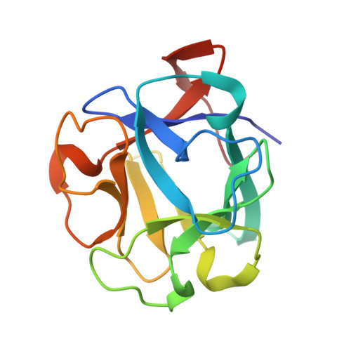
5VBK - PubMed Abstract:
Lectins play crucial roles for innate immune responses in invertebrates by recognizing and eliminating pathogens. In this study, a lectin from the mussel Mytilus californianus (MCL) was identified and characterized. The lectin was purified by affinity chromatography in α-lactose-agarose resin showing an experimental molecular mass of 18000 Da as determined by SDS-PAGE and MALDI-TOF mass spectrometry. It was specific for binding d-galactose and N-Acetyl-d-galactosamine that contained carbohydrate moieties that were also inhibited by melibiose and raffinose. It had the ability to agglutinate all types of human erythrocytes, as well as rabbit red blood cells. Circular dichroism analyzes have indicated that this lectin possessed an α/β fold with a predominance of β structures. This was consistent with the structure of the protein that was determined by the X-ray diffraction techniques. MCL was crystallized in the space group C2 1 and it diffracted to 1.79 Å resolution. Two monomers were found in the asymmetric unit and they formed dimers in solution. The protein has shown to be a member of the β-trefoil family, with three sugar binding sites per monomer. In accord with fluorescence-based thermal shift assays, we observed that the MCL T m increased about 10 °C in the presence of galactose. Furthermore, we have determined the complete amino acid sequence by cDNA sequencing. The gene had two ORF2 proteins, one resulting in a 180 residue protein with a theoretical molecular mass of 20227 Da, and another resulting in a 150 residue protein with a theoretical molecular mass of 16911 Da. The difference between the theoretical and experimental values was due to the presence of a glycosylation that was observed by the glycosylation assay. A positive microbial agglutination and a growth inhibition activity were observed against Gram-negative and Gram-positive bacteria. The M. californianus lectin is the fourth member of the recently proposed new family of lectins that have been reported to date, occurring only in mollusks belonging to the family Mytilidae. It is the first member to be glycosylated and with a strong tendency to form large oligomers.
- Departamento de Química de Biomacromoléculas, Instituto de Química, Universidad Nacional Autónoma de México. Circuito Exterior, Ciudad Universitaria, Coyoacán, Cd. Mx. C.P. 04510, Mexico.
Organizational Affiliation: