5VB9 | pdb_00005vb9

IL-17A in complex with peptide


Experimental Data Snapshot

  • Method: X-RAY DIFFRACTION
  • Resolution: 1.70 Å
  • R-Value Free: 
    0.211 (Depositor), 0.211 (DCC) 
  • R-Value Work: 
    0.182 (Depositor), 0.187 (DCC) 
  • R-Value Observed: 
    0.183 (Depositor) 

wwPDB Validation 3D Report Full Report

Validation slider image for 5VB9

This is version 1.1 of the entry. See complete history

Literature

Utilization of peptide phage display to investigate hotspots on IL-17A and what it means for drug discovery.

Ting, J.P.Tung, F.Antonysamy, S.Wasserman, S.Jones, S.B.Zhang, F.F.Espada, A.Broughton, H.Chalmers, M.J.Woodman, M.E.Bina, H.A.Dodge, J.A.Benach, J.Zhang, A.Groshong, C.Manglicmot, D.Russell, M.Afshar, S.

(2018) PLoS One 13: e0190850-e0190850

  • DOI: https://doi.org/10.1371/journal.pone.0190850
  • Primary Citation Related Structures: 
    5VB9

  • PubMed Abstract: 

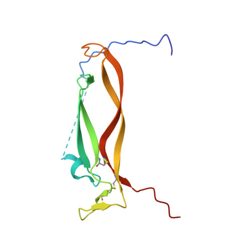
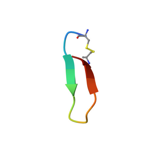
    To date, IL-17A antibodies remain the only therapeutic approach to correct the abnormal activation of the IL-17A/IL-17R signaling complex. Why is it that despite the remarkable success of IL-17 antibodies, there is no small molecule antagonist of IL-17A in the clinic? Here we offer a unique approach to address this question. In order to understand the interaction of IL-17A with its receptor, we combined peptide discovery using phage display with HDX, crystallography, and functional assays to map and characterize hot regions that contribute to most of the energetics of the IL-17A/IL-17R interaction. These functional maps are proposed to serve as a guide to aid in the development of small molecules that bind to IL-17A and block its interaction with IL-17RA.


  • Organizational Affiliation
    • Department of protein Engineering, Eli Lilly Biotechnology Center, San Diego, California, United States of America.

Macromolecule Content 

  • Total Structure Weight: 31.41 kDa 
  • Atom Count: 2,267 
  • Modeled Residue Count: 239 
  • Deposited Residue Count: 268 
  • Unique protein chains: 2

Macromolecules

Find similar proteins by:|  3D Structure
Entity ID: 1
MoleculeChains  Sequence LengthOrganismDetailsImage
Interleukin-17A
A, B
119Homo sapiensMutation(s): 0 
Gene Names: IL17ACTLA8IL17
UniProt & NIH Common Fund Data Resources
Find proteins for Q16552 (Homo sapiens)
Explore Q16552 
Go to UniProtKB:  Q16552
PHAROS:  Q16552
GTEx:  ENSG00000112115 
Entity Groups
Sequence Clusters30% Identity50% Identity70% Identity90% Identity95% Identity100% Identity
UniProt GroupQ16552
Sequence Annotations
Expand
Reference Sequence
Find similar proteins by:  Sequence   |   3D Structure  
Entity ID: 2
MoleculeChains  Sequence LengthOrganismDetailsImage
Peptide inhibitor
C, D
15synthetic constructMutation(s): 0 

Experimental Data & Validation

Experimental Data

  • Method: X-RAY DIFFRACTION
  • Resolution: 1.70 Å
  • R-Value Free:  0.211 (Depositor), 0.211 (DCC) 
  • R-Value Work:  0.182 (Depositor), 0.187 (DCC) 
  • R-Value Observed: 0.183 (Depositor) 
Space Group: P 21 21 2
Unit Cell:
Length ( Å )Angle ( ˚ )
a = 36.771α = 90
b = 55.122β = 90
c = 143.953γ = 90
Software Package:
Software NamePurpose
MOSFLMdata reduction
SCALAdata scaling
PHASERphasing
BUSTERrefinement
PDB_EXTRACTdata extraction

Structure Validation

View Full Validation Report


Entry History 

Revision History  (Full details and data files)

  • Version 1.0: 2018-02-14
    Type: Initial release
  • Version 1.1: 2024-10-30
    Changes: Data collection, Database references, Structure summary