5V4H | pdb_00005v4h

Ruthenium(II)(cymene)(chlorido)2-lysozyme adduct formed when ruthenium(II)(cymene)(bromido)2 underwent ligand exchange, resulting in one binding site


Experimental Data Snapshot

  • Method: X-RAY DIFFRACTION
  • Resolution: 1.22 Å
  • R-Value Free: 
    0.190 (Depositor), 0.192 (DCC) 
  • R-Value Work: 
    0.154 (Depositor), 0.154 (DCC) 
  • R-Value Observed: 
    0.156 (Depositor) 

Starting Model: experimental
View more details

wwPDB Validation 3D Report Full Report

Validation slider image for 5V4H

Ligand Structure Quality Assessment 


This is version 1.4 of the entry. See complete history

Literature

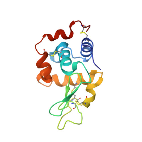
The metalation of hen egg white lysozyme impacts protein stability as shown by ion mobility mass spectrometry, differential scanning calorimetry, and X-ray crystallography.

Sullivan, M.P.Groessl, M.Meier, S.M.Kingston, R.L.Goldstone, D.C.Hartinger, C.G.

(2017) Chem Commun (Camb) 53: 4246-4249

  • DOI: https://doi.org/10.1039/c6cc10150j
  • Primary Citation Related Structures: 
    5V4G, 5V4H, 5V4I

  • PubMed Abstract: 

    Metalation of hen egg white lysozyme (HEWL) with organometallics was studied with physicochemical methods in solid state, solution and the gas phase. While metalation did not affect the crystal structure of HEWL significantly, protein destabilisation was detected in gas phase and solution.


  • Organizational Affiliation
    • School of Chemical Sciences, University of Auckland, Private Bag 92019, Auckland, 1142, New Zealand. c.hartinger@auckland.ac.nz.

Macromolecule Content 

  • Total Structure Weight: 14.66 kDa 
  • Atom Count: 1,163 
  • Modeled Residue Count: 129 
  • Deposited Residue Count: 129 
  • Unique protein chains: 1

Macromolecules

Find similar proteins by:|  3D Structure
Entity ID: 1
MoleculeChains  Sequence LengthOrganismDetailsImage
Lysozyme C129Gallus gallusMutation(s): 0 
EC: 3.2.1.17
UniProt
Find proteins for P00698 (Gallus gallus)
Explore P00698 
Go to UniProtKB:  P00698
Entity Groups
Sequence Clusters30% Identity50% Identity70% Identity90% Identity95% Identity100% Identity
UniProt GroupP00698
Sequence Annotations
Expand
Reference Sequence

Small Molecules

Experimental Data & Validation

Experimental Data

  • Method: X-RAY DIFFRACTION
  • Resolution: 1.22 Å
  • R-Value Free:  0.190 (Depositor), 0.192 (DCC) 
  • R-Value Work:  0.154 (Depositor), 0.154 (DCC) 
  • R-Value Observed: 0.156 (Depositor) 
Space Group: P 43 21 2
Unit Cell:
Length ( Å )Angle ( ˚ )
a = 80.32α = 90
b = 80.32β = 90
c = 36.59γ = 90
Software Package:
Software NamePurpose
REFMACrefinement
iMOSFLMdata reduction
Aimlessdata scaling
PHASERphasing
Cootmodel building
PDB_EXTRACTdata extraction

Structure Validation

View Full Validation Report


Ligand Structure Quality Assessment 


Entry History 

Deposition Data

Revision History  (Full details and data files)

  • Version 1.0: 2017-04-05
    Type: Initial release
  • Version 1.1: 2017-04-12
    Changes: Database references
  • Version 1.2: 2017-04-19
    Changes: Database references
  • Version 1.3: 2023-10-04
    Changes: Data collection, Database references, Derived calculations, Refinement description
  • Version 1.4: 2024-11-13
    Changes: Structure summary