


Crystal Structure of the Group II Truncated Hemoglobin from Bacillus Anthracis
Varnado, C.L., SoRelle, E., Soman, J., Olson, J.S.To be published.
Experimental Data Snapshot
Entity ID: 1 | |||||
|---|---|---|---|---|---|
| Molecule | Chains | Sequence Length | Organism | Details | Image |
| Globin | 132 | Bacillus anthracis | Mutation(s): 1 Gene Names: yjbI, GBAA_1209, BASH2_04647, BVB96_06425 | 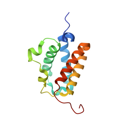 | |
UniProt | |||||
Entity Groups | |||||
| Sequence Clusters | 30% Identity50% Identity70% Identity90% Identity95% Identity100% Identity | ||||
| UniProt Group | A0A6L7H965 | ||||
Sequence AnnotationsExpand | |||||
Reference Sequence | |||||
| Ligands 3 Unique | |||||
|---|---|---|---|---|---|
| ID | Chains | Name / Formula / InChI Key | 2D Diagram | 3D Interactions | |
| HEM Download:Ideal Coordinates CCD File | C [auth A], G [auth B] | PROTOPORPHYRIN IX CONTAINING FE C34 H32 Fe N4 O4 KABFMIBPWCXCRK-RGGAHWMASA-L |  | ||
| SO4 Download:Ideal Coordinates CCD File | E [auth A], F [auth A], I [auth B] | SULFATE ION O4 S QAOWNCQODCNURD-UHFFFAOYSA-L |  | ||
| CYN Download:Ideal Coordinates CCD File | D [auth A], H [auth B] | CYANIDE ION C N XFXPMWWXUTWYJX-UHFFFAOYSA-N |  | ||
| Length ( Å ) | Angle ( ˚ ) |
|---|---|
| a = 49.449 | α = 90 |
| b = 122.303 | β = 90 |
| c = 111.494 | γ = 90 |
| Software Name | Purpose |
|---|---|
| PHENIX | refinement |
| d*TREK | data scaling |
| d*TREK | data reduction |
| PDB_EXTRACT | data extraction |
| PHENIX | phasing |


