5V1B | pdb_00005v1b

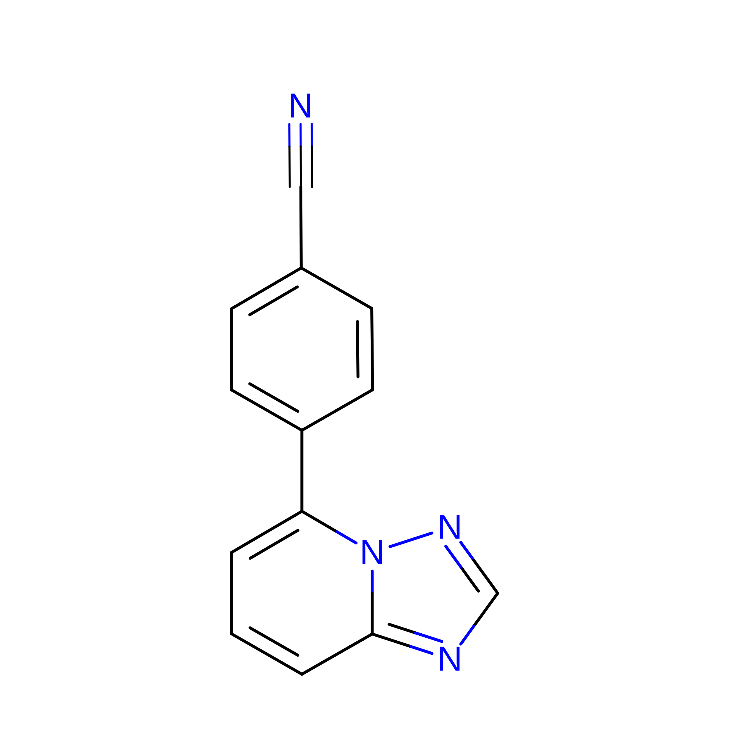
Structure of PHD1 in complex with 1,2,4-Triazolo-[1,5-a]pyridine


Experimental Data Snapshot

  • Method: X-RAY DIFFRACTION
  • Resolution: 2.49 Å
  • R-Value Free: 
    0.236 (Depositor), 0.242 (DCC) 
  • R-Value Work: 
    0.189 (Depositor), 0.197 (DCC) 
  • R-Value Observed: 
    0.192 (Depositor) 

wwPDB Validation 3D Report Full Report

Validation slider image for 5V1B

Ligand Structure Quality Assessment 


This is version 1.2 of the entry. See complete history. 

Literature

1,2,4-Triazolo-[1,5-a]pyridine HIF Prolylhydroxylase Domain-1 (PHD-1) Inhibitors With a Novel Monodentate Binding Interaction.

Ahmed, S., Ayscough, A., Barker, G.R., Canning, H.E., Davenport, R., Downham, R., Harrison, D., Jenkins, K., Kinsella, N., Livermore, D.G., Wright, S., Ivetac, A.D., Skene, R., Wilkens, S.J., Webster, N.A., Hendrick, A.G.

(2017) J Med Chem 60: 5663-5672

  • DOI: https://doi.org/10.1021/acs.jmedchem.7b00352
  • Primary Citation Related Structures: 
    5V18, 5V1B

  • PubMed Abstract: 

    Herein we describe the identification of 4-{[1,2,4]triazolo[1,5-a]pyridin-5-yl}benzonitrile-based inhibitors of the hypoxia-inducible factor prolylhydroxylase domain-1 (PHD-1) enzyme. These inhibitors were shown to possess a novel binding mode by X-ray crystallography, in which the triazolo N1 atom coordinates in a hitherto unreported monodentate interaction with the active site Fe 2+ ion, while the benzonitrile group accepts a hydrogen-bonding interaction from the side chain residue of Asn315. Further optimization led to potent PHD-1 inhibitors with good physicochemical and pharmacokinetic properties.


  • Organizational Affiliation: 
    • Department of Computational Sciences and Crystallography, Takeda California Inc. , 10410 Science Center Dr., San Diego, California 92121, United States.

Macromolecule Content 

  • Total Structure Weight: 27.03 kDa 
  • Atom Count: 1,891 
  • Modeled Residue Count: 226 
  • Deposited Residue Count: 240 
  • Unique protein chains: 1

Macromolecules

Find similar proteins by:|  3D Structure
Entity ID: 1
MoleculeChains  Sequence LengthOrganismDetailsImage
Egl nine homolog 2240Homo sapiensMutation(s): 0 
Gene Names: EGLN2, EIT6
EC: 1.14.11.29 (PDB Primary Data), 1.14.11 (UniProt)
UniProt & NIH Common Fund Data Resources
Find proteins for Q96KS0 (Homo sapiens)
Explore Q96KS0 
Go to UniProtKB:  Q96KS0
PHAROS:  Q96KS0
GTEx:  ENSG00000269858 
Entity Groups
Sequence Clusters30% Identity50% Identity70% Identity90% Identity95% Identity100% Identity
UniProt GroupQ96KS0
Sequence Annotations
Expand
Reference Sequence

Experimental Data & Validation

Experimental Data

  • Method: X-RAY DIFFRACTION
  • Resolution: 2.49 Å
  • R-Value Free:  0.236 (Depositor), 0.242 (DCC) 
  • R-Value Work:  0.189 (Depositor), 0.197 (DCC) 
  • R-Value Observed: 0.192 (Depositor) 
Space Group: C 2 2 21
Unit Cell:
Length ( Å )Angle ( ˚ )
a = 66.717α = 90
b = 112.082β = 90
c = 66.223γ = 90
Software Package:
Software NamePurpose
SCALEPACKdata scaling
REFMACrefinement
PDB_EXTRACTdata extraction
DENZOdata reduction
PHASERphasing

Structure Validation

View Full Validation Report


Ligand Structure Quality Assessment 


Entry History 

Deposition Data

  • Released Date: 2017-06-21 
  • Deposition Author(s): Skene, R.J.

Revision History  (Full details and data files)

  • Version 1.0: 2017-06-21
    Type: Initial release
  • Version 1.1: 2017-07-26
    Changes: Database references
  • Version 1.2: 2024-03-06
    Changes: Data collection, Database references, Derived calculations