5V12 | pdb_00005v12

Crystal structure of Carbon Sulfoxide lyase, Egt2 Y134F with sulfenic acid intermediate


Experimental Data Snapshot

  • Method: X-RAY DIFFRACTION
  • Resolution: 2.45 Å
  • R-Value Free: 
    0.272 (Depositor), 0.273 (DCC) 
  • R-Value Work: 
    0.222 (Depositor), 0.223 (DCC) 
  • R-Value Observed: 
    0.223 (Depositor) 

Starting Model: experimental
View more details

wwPDB Validation 3D Report Full Report

Validation slider image for 5V12

Ligand Structure Quality Assessment 


This is version 3.0 of the entry. See complete history

Literature

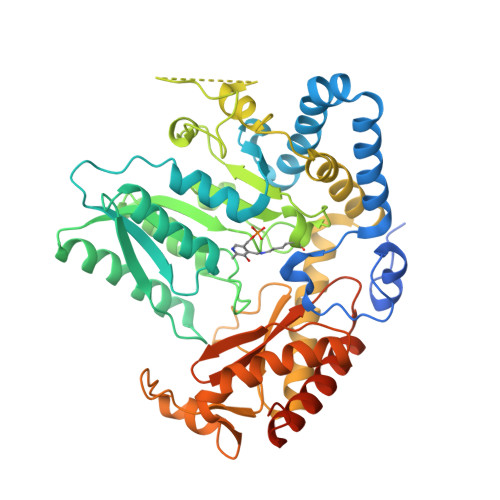
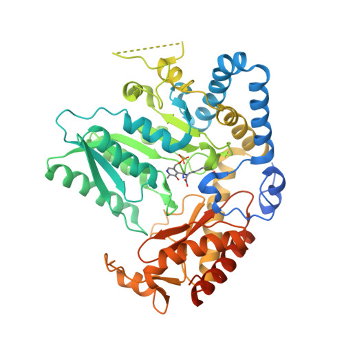
Snapshots of C-S Cleavage in Egt2 Reveals Substrate Specificity and Reaction Mechanism.

Irani, S.Naowarojna, N.Tang, Y.Kathuria, K.R.Wang, S.Dhembi, A.Lee, N.Yan, W.Lyu, H.Costello, C.E.Liu, P.Zhang, Y.J.

(2018) Cell Chem Biol 25: 519-529.e4

  • DOI: https://doi.org/10.1016/j.chembiol.2018.02.002
  • Primary Citation Related Structures: 
    5UTS, 5V12, 5V1X

  • PubMed Abstract: 

    Sulfur incorporation in the biosynthesis of ergothioneine, a histidine thiol derivative, differs from other well-characterized transsulfurations. A combination of a mononuclear non-heme iron enzyme-catalyzed oxidative C-S bond formation and a subsequent pyridoxal 5'-phosphate (PLP)-mediated C-S lyase reaction leads to the net transfer of a sulfur atom from a cysteine to a histidine. In this study, we structurally and mechanistically characterized a PLP-dependent C-S lyase Egt2, which mediates the sulfoxide C-S bond cleavage in ergothioneine biosynthesis. A cation-π interaction between substrate and enzyme accounts for Egt2's preference of sulfoxide over thioether as a substrate. Using mutagenesis and structural biology, we captured three distinct states of the Egt2 C-S lyase reaction cycle, including a labile sulfenic intermediate captured in Egt2 crystals. Chemical trapping and high-resolution mass spectrometry were used to confirm the involvement of the sulfenic acid intermediate in Egt2 catalysis.


  • Organizational Affiliation
    • Department of Chemical Engineering, University of Texas at Austin, Austin, TX 78712, USA.

Macromolecule Content 

  • Total Structure Weight: 452.36 kDa 
  • Atom Count: 28,363 
  • Modeled Residue Count: 3,502 
  • Deposited Residue Count: 4,008 
  • Unique protein chains: 2

Macromolecules

Find similar proteins by:|  3D Structure
Entity ID: 1
MoleculeChains  Sequence LengthOrganismDetailsImage
Hercynylcysteine sulfoxide lyase501Neurospora crassa OR74AMutation(s): 0 
Gene Names: egt-2NCU11365
EC: 4.4.1 (PDB Primary Data), 4.4.1.36 (UniProt)
UniProt
Find proteins for A7UX13 (Neurospora crassa (strain ATCC 24698 / 74-OR23-1A / CBS 708.71 / DSM 1257 / FGSC 987))
Explore A7UX13 
Go to UniProtKB:  A7UX13
Entity Groups
Sequence Clusters30% Identity50% Identity70% Identity90% Identity95% Identity100% Identity
UniProt GroupA7UX13
Sequence Annotations
Expand
Reference Sequence
Find similar proteins by:|  3D Structure
Entity ID: 2
MoleculeChains  Sequence LengthOrganismDetailsImage
Hercynylcysteine sulfoxide lyaseE [auth C]501Neurospora crassa OR74AMutation(s): 0 
Gene Names: egt-2NCU11365
EC: 4.4.1 (PDB Primary Data), 4.4.1.36 (UniProt)
UniProt
Find proteins for A7UX13 (Neurospora crassa (strain ATCC 24698 / 74-OR23-1A / CBS 708.71 / DSM 1257 / FGSC 987))
Explore A7UX13 
Go to UniProtKB:  A7UX13
Entity Groups
Sequence Clusters30% Identity50% Identity70% Identity90% Identity95% Identity100% Identity
UniProt GroupA7UX13
Sequence Annotations
Expand
Reference Sequence

Small Molecules

Ligands 2 Unique
IDChains Name / Formula / InChI Key2D Diagram3D Interactions
8QJ

Query on 8QJ


Download:Ideal Coordinates CCD File
K [auth F],
P [auth A]
(1S)-1-carboxy-2-[2-(hydroxysulfanyl)-1H-imidazol-4-yl]-N,N,N-trimethylethan-1-aminium
C9 H16 N3 O3 S
ZLPHHLCGSBYNCO-ZETCQYMHSA-O
FMT

Query on FMT


Download:Ideal Coordinates CCD File
I [auth E]
J [auth G]
L [auth F]
M [auth H]
N [auth C]
I [auth E],
J [auth G],
L [auth F],
M [auth H],
N [auth C],
O [auth D],
Q [auth A],
R [auth B]
FORMIC ACID
C H2 O2
BDAGIHXWWSANSR-UHFFFAOYSA-N
Modified Residues  2 Unique
IDChains TypeFormula2D DiagramParent
LLP
Query on LLP
A [auth E]
B [auth G]
C [auth F]
D [auth H]
F [auth D]
A [auth E],
B [auth G],
C [auth F],
D [auth H],
F [auth D],
G [auth A],
H [auth B]
L-PEPTIDE LINKINGC14 H22 N3 O7 PLYS
EXA
Query on EXA
E [auth C]PEPTIDE LINKINGC17 H27 N4 O9 PLYS

Experimental Data & Validation

Experimental Data

  • Method: X-RAY DIFFRACTION
  • Resolution: 2.45 Å
  • R-Value Free:  0.272 (Depositor), 0.273 (DCC) 
  • R-Value Work:  0.222 (Depositor), 0.223 (DCC) 
  • R-Value Observed: 0.223 (Depositor) 
Space Group: P 1 21 1
Unit Cell:
Length ( Å )Angle ( ˚ )
a = 104.021α = 90
b = 195.158β = 91.47
c = 110.1γ = 90
Software Package:
Software NamePurpose
PHENIXrefinement
HKL-2000data reduction
HKL-2000data scaling

Structure Validation

View Full Validation Report


Ligand Structure Quality Assessment 


Entry History 

Deposition Data

Revision History  (Full details and data files)

  • Version 1.0: 2018-03-07
    Type: Initial release
  • Version 1.1: 2019-03-20
    Changes: Data collection, Database references, Structure summary
  • Version 2.0: 2023-10-04
    Changes: Atomic model, Data collection, Database references, Derived calculations, Polymer sequence, Refinement description, Source and taxonomy, Structure summary
  • Version 2.1: 2023-11-15
    Changes: Data collection
  • Version 2.2: 2023-12-06
    Changes: Data collection
  • Version 3.0: 2024-05-01
    Changes: Derived calculations, Non-polymer description, Structure summary