5UYX | pdb_00005uyx

Structure of Human T-complex protein 1 subunit epsilon (CCT5)


Experimental Data Snapshot

  • Method: X-RAY DIFFRACTION
  • Resolution: 3.50 Å
  • R-Value Free: 
    0.318 (Depositor), 0.320 (DCC) 
  • R-Value Work: 
    0.269 (Depositor), 0.270 (DCC) 
  • R-Value Observed: 
    0.271 (Depositor) 

Starting Model: experimental
View more details

wwPDB Validation   3D Report Full Report


Ligand Structure Quality Assessment 

Created with Raphaël 2.3.0Worse 01 BetterLigand structure goodness of fit to experimental dataBest fitted ADPClick on this verticalbar to view details

This is version 1.1 of the entry. See complete history


Literature

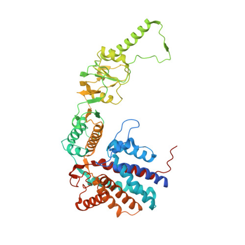
Structure of the human TRiC/CCT Subunit 5 associated with hereditary sensory neuropathy.

Pereira, J.H.McAndrew, R.P.Sergeeva, O.A.Ralston, C.Y.King, J.A.Adams, P.D.

(2017) Sci Rep 7: 3673-3673

  • DOI: https://doi.org/10.1038/s41598-017-03825-3
  • Primary Citation of Related Structures:  
    5UYX, 5UYZ

  • PubMed Abstract: 

    The human chaperonin TRiC consists of eight non-identical subunits, and its protein-folding activity is critical for cellular health. Misfolded proteins are associated with many human diseases, such as amyloid diseases, cancer, and neuropathies, making TRiC a potential therapeutic target. A detailed structural understanding of its ATP-dependent folding mechanism and substrate recognition is therefore of great importance. Of particular health-related interest is the mutation Histidine 147 to Arginine (H147R) in human TRiC subunit 5 (CCT5), which has been associated with hereditary sensory neuropathy. In this paper, we describe the crystal structures of CCT5 and the CCT5-H147R mutant, which provide important structural information for this vital protein-folding machine in humans. This first X-ray crystallographic study of a single human CCT subunit in the context of a hexadecameric complex can be expanded in the future to the other 7 subunits that form the TRiC complex.


  • Organizational Affiliation

    Molecular Biophysics and Integrated Bioimaging Division, Lawrence Berkeley National Laboratory, Berkeley, CA, 94720, USA.


Macromolecules
Find similar proteins by:  (by identity cutoff)  |  3D Structure
Entity ID: 1
MoleculeChains Sequence LengthOrganismDetailsImage
T-complex protein 1 subunit epsilon
A, B, C, D
541Homo sapiensMutation(s): 0 
Gene Names: CCT5CCTEKIAA0098
UniProt & NIH Common Fund Data Resources
Find proteins for P48643 (Homo sapiens)
Explore P48643 
Go to UniProtKB:  P48643
PHAROS:  P48643
GTEx:  ENSG00000150753 
Entity Groups  
Sequence Clusters30% Identity50% Identity70% Identity90% Identity95% Identity100% Identity
UniProt GroupP48643
Sequence Annotations
Expand
  • Reference Sequence
Experimental Data & Validation

Experimental Data

  • Method: X-RAY DIFFRACTION
  • Resolution: 3.50 Å
  • R-Value Free:  0.318 (Depositor), 0.320 (DCC) 
  • R-Value Work:  0.269 (Depositor), 0.270 (DCC) 
  • R-Value Observed: 0.271 (Depositor) 
Space Group: P 4 21 2
Unit Cell:
Length ( Å )Angle ( ˚ )
a = 204.71α = 90
b = 204.71β = 90
c = 162.95γ = 90
Software Package:
Software NamePurpose
PHENIXrefinement
xia2data reduction
xia2data scaling
PHASERphasing

Structure Validation

View Full Validation Report


Ligand Structure Quality Assessment 

Created with Raphaël 2.3.0Worse 01 BetterLigand structure goodness of fit to experimental dataBest fitted ADPClick on this verticalbar to view details

Entry History 

Deposition Data

Revision History  (Full details and data files)

  • Version 1.0: 2017-07-05
    Type: Initial release
  • Version 1.1: 2023-10-04
    Changes: Data collection, Database references, Refinement description