5UXA | pdb_00005uxa

Crystal structure of macrolide 2'-phosphotransferase MphB from Escherichia coli


Experimental Data Snapshot

  • Method: X-RAY DIFFRACTION
  • Resolution: 1.95 Å
  • R-Value Free: 
    0.201 (Depositor), 0.198 (DCC) 
  • R-Value Work: 
    0.151 (Depositor), 0.150 (DCC) 
  • R-Value Observed: 
    0.155 (Depositor) 

wwPDB Validation 3D Report Full Report

Validation slider image for 5UXA

This is version 2.2 of the entry. See complete history

Literature

The evolution of substrate discrimination in macrolide antibiotic resistance enzymes.

Pawlowski, A.C.Stogios, P.J.Koteva, K.Skarina, T.Evdokimova, E.Savchenko, A.Wright, G.D.

(2018) Nat Commun 9: 112-112

  • DOI: https://doi.org/10.1038/s41467-017-02680-0
  • Primary Citation Related Structures: 
    5UXA, 5UXB, 5UXC, 5UXD

  • PubMed Abstract: 

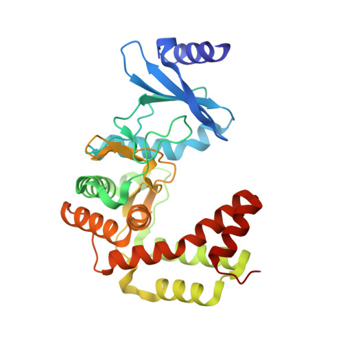
    The production of antibiotics by microbes in the environment and their use in medicine and agriculture select for existing and emerging resistance. To address this inevitability, prudent development of antibiotic drugs requires careful consideration of resistance evolution. Here, we identify the molecular basis for expanded substrate specificity in MphI, a macrolide kinase (Mph) that does not confer resistance to erythromycin, in contrast to other known Mphs. Using a combination of phylogenetics, drug-resistance phenotypes, and in vitro enzyme assays, we find that MphI and MphK phosphorylate erythromycin poorly resulting in an antibiotic-sensitive phenotype. Using likelihood reconstruction of ancestral sequences and site-saturation combinatorial mutagenesis, supported by Mph crystal structures, we determine that two non-obvious mutations in combination expand the substrate range. This approach should be applicable for studying the functional evolution of any antibiotic resistance enzyme and for evaluating the evolvability of resistance enzymes to new generations of antibiotic scaffolds.


  • Organizational Affiliation
    • Michael G. DeGroote Institute for Infectious Disease Research and the Department of Biochemistry and Biomedical Sciences, McMaster University, Hamilton, L8S 4L8, ON, Canada.

Macromolecule Content 

  • Total Structure Weight: 34.65 kDa 
  • Atom Count: 2,904 
  • Modeled Residue Count: 301 
  • Deposited Residue Count: 302 
  • Unique protein chains: 1

Macromolecules

Find similar proteins by:|  3D Structure
Entity ID: 1
MoleculeChains  Sequence LengthOrganismDetailsImage
Macrolide 2'-phosphotransferase II302Escherichia coliMutation(s): 0 
Gene Names: mphBpO103_99
UniProt
Find proteins for O32553 (Escherichia coli)
Explore O32553 
Go to UniProtKB:  O32553
Entity Groups
Sequence Clusters30% Identity50% Identity70% Identity90% Identity95% Identity100% Identity
UniProt GroupO32553
Sequence Annotations
Expand
Reference Sequence

Experimental Data & Validation

Experimental Data

  • Method: X-RAY DIFFRACTION
  • Resolution: 1.95 Å
  • R-Value Free:  0.201 (Depositor), 0.198 (DCC) 
  • R-Value Work:  0.151 (Depositor), 0.150 (DCC) 
  • R-Value Observed: 0.155 (Depositor) 
Space Group: C 2 2 21
Unit Cell:
Length ( Å )Angle ( ˚ )
a = 67.276α = 90
b = 116.684β = 90
c = 92.574γ = 90
Software Package:
Software NamePurpose
PHENIXrefinement
HKL-3000data reduction
HKL-3000data scaling
PHENIXphasing
PHENIXmodel building
Cootmodel building

Structure Validation

View Full Validation Report


Entry History 

& Funding Information

Deposition Data


Funding OrganizationLocationGrant Number
National Institutes of Health/National Institute Of Allergy and Infectious Diseases (NIH/NIAID)United StatesHHSCN27220120026C

Revision History  (Full details and data files)

  • Version 1.0: 2017-06-28
    Type: Initial release
  • Version 1.1: 2017-09-13
    Changes: Author supporting evidence
  • Version 1.2: 2018-01-31
    Changes: Experimental preparation
  • Version 2.0: 2018-02-21
    Changes: Database references, Polymer sequence
  • Version 2.1: 2019-12-11
    Changes: Author supporting evidence
  • Version 2.2: 2024-03-06
    Changes: Data collection, Database references, Derived calculations