5UR0 | pdb_00005ur0

Crystallographic structure of glyceraldehyde-3-phosphate dehydrogenase from Naegleria gruberi


Experimental Data Snapshot

  • Method: X-RAY DIFFRACTION
  • Resolution: 1.94 Å
  • R-Value Free: 
    0.198 (Depositor), 0.197 (DCC) 
  • R-Value Work: 
    0.154 (Depositor), 0.155 (DCC) 
  • R-Value Observed: 
    0.154 (Depositor) 

Starting Model: experimental
View more details

wwPDB Validation 3D Report Full Report

Validation slider image for 5UR0

Ligand Structure Quality Assessment 


This is version 1.7 of the entry. See complete history

Literature

Structural studies of glyceraldehyde-3-phosphate dehydrogenase from Naegleria gruberi, the first one from phylum Percolozoa.

Machado, A.T.P.Silva, M.Iulek, J.

(2018) Biochim Biophys Acta 1866: 581-588

  • DOI: https://doi.org/10.1016/j.bbapap.2018.02.006
  • Primary Citation Related Structures: 
    5UR0

  • PubMed Abstract: 

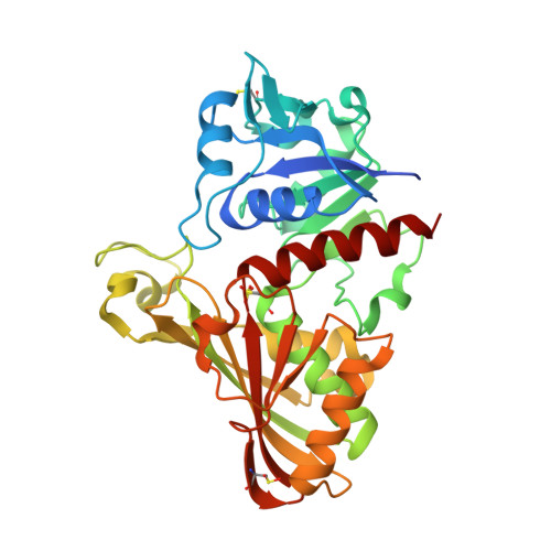
    Naegleria gruberi is a free life amoeba believed to have more than one billion years of existence; it is not pathogenic and had its genome sequenced, which revealed a high complexity in the metabolic pathways. This paper presents the experimental structure of GAPDH from N. gruberi, the first one belonging to the phylum Percolozoa, comparisons to structures from various species and molecular dynamics studies of some particular features. The final refined structure presents R cryst  = 15.54% and R free  = 19.84%. The catalytic domain formed by residues 134 to 313 is highly conserved, as expected, with the exception of Asn145, present only in NgGAPDH, while the other GAPDHs present either Ser or Thr on the corresponding position. Molecular dynamics analysis revealed that Asn145 has correlated motions with residues Ala123, Thr125 and Pro126 that belong to what was called "bonded loop". NgGAPDH residue Met35 presents an extended side chain, closer to the cofactor adenine ring than corresponding (different) residues and conformations found in some parasitic protozoa and the human GAPDHs. The enzyme was previously reported to present positive cooperativity, which is hypothesized to be related to certain atom distances.


  • Organizational Affiliation
    • Chemistry, State University of Ponta Grossa, Ponta Grossa 84030-900, Brazil.

Macromolecule Content 

  • Total Structure Weight: 157.28 kDa 
  • Atom Count: 11,786 
  • Modeled Residue Count: 1,341 
  • Deposited Residue Count: 1,400 
  • Unique protein chains: 1

Macromolecules

Find similar proteins by:|  3D Structure
Entity ID: 1
MoleculeChains  Sequence LengthOrganismDetailsImage
Glyceraldehyde-3-phosphate dehydrogenase
A, B, C, D
349Naegleria gruberiMutation(s): 1 
Gene Names: NAEGRDRAFT_53883
EC: 1.2.1.12
UniProt
Find proteins for D2W142 (Naegleria gruberi)
Explore D2W142 
Go to UniProtKB:  D2W142
Entity Groups
Sequence Clusters30% Identity50% Identity70% Identity90% Identity95% Identity100% Identity
UniProt GroupD2W142
Sequence Annotations
Expand
Reference Sequence

Small Molecules

Ligands 3 Unique
IDChains Name / Formula / InChI Key2D Diagram3D Interactions
NAD

Query on NAD


Download:Ideal Coordinates CCD File
E [auth A],
H [auth B],
J [auth C],
L [auth D]
NICOTINAMIDE-ADENINE-DINUCLEOTIDE
C21 H27 N7 O14 P2
BAWFJGJZGIEFAR-NNYOXOHSSA-N
PGE

Query on PGE


Download:Ideal Coordinates CCD File
F [auth A]TRIETHYLENE GLYCOL
C6 H14 O4
ZIBGPFATKBEMQZ-UHFFFAOYSA-N
NA

Query on NA


Download:Ideal Coordinates CCD File
G [auth A],
I [auth B],
K [auth C],
M [auth D]
SODIUM ION
Na
FKNQFGJONOIPTF-UHFFFAOYSA-N
Modified Residues  3 Unique
IDChains TypeFormula2D DiagramParent
CME
Query on CME
A, B, C, D
L-PEPTIDE LINKINGC5 H11 N O3 S2CYS
CSD
Query on CSD
A, B, C, D
L-PEPTIDE LINKINGC3 H7 N O4 SCYS
OCS
Query on OCS
A, B, C, D
L-PEPTIDE LINKINGC3 H7 N O5 SCYS

Experimental Data & Validation

Experimental Data

  • Method: X-RAY DIFFRACTION
  • Resolution: 1.94 Å
  • R-Value Free:  0.198 (Depositor), 0.197 (DCC) 
  • R-Value Work:  0.154 (Depositor), 0.155 (DCC) 
  • R-Value Observed: 0.154 (Depositor) 
Space Group: P 1 21 1
Unit Cell:
Length ( Å )Angle ( ˚ )
a = 83.743α = 90
b = 94.552β = 99.96
c = 90.932γ = 90
Software Package:
Software NamePurpose
PHENIXrefinement
XDSdata processing
XSCALEdata scaling
PHASERphasing
Cootmodel building

Structure Validation

View Full Validation Report


Ligand Structure Quality Assessment 


Entry History 

& Funding Information

Deposition Data


Funding OrganizationLocationGrant Number
Brazilian National Council for Scientific and Technological Development (CNPq)Brazil573607/2008-7

Revision History  (Full details and data files)

  • Version 1.0: 2018-03-14
    Type: Initial release
  • Version 1.1: 2018-03-28
    Changes: Data collection, Database references
  • Version 1.2: 2018-04-18
    Changes: Data collection
  • Version 1.3: 2019-04-17
    Changes: Author supporting evidence, Data collection
  • Version 1.4: 2020-01-08
    Changes: Author supporting evidence
  • Version 1.5: 2023-10-04
    Changes: Data collection, Database references, Derived calculations, Refinement description
  • Version 1.6: 2023-11-15
    Changes: Data collection
  • Version 1.7: 2024-11-13
    Changes: Structure summary