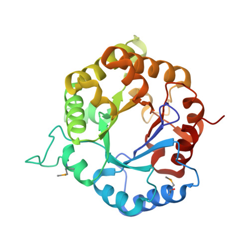
X-ray structure of a putative triosephosphate isomerase from Toxoplasma gondii ME49
Filippova, E.V., Wawrzak, Z., Minasov, G., Cardona-Correa, A., Bishop, B., Anderson, W.F., Ngo, H., Center for Structural Genomics of Infectious Diseases (CSGID)To be published.