K-Ras Populates Conformational States Differently from Its Isoform H-Ras and Oncogenic Mutant K-RasG12D.
Parker, J.A., Volmar, A.Y., Pavlopoulos, S., Mattos, C.(2018) Structure 26: 810
- PubMed: 29706533 Search on PubMed
- DOI: https://doi.org/10.1016/j.str.2018.03.018
- Primary Citation Related Structures:
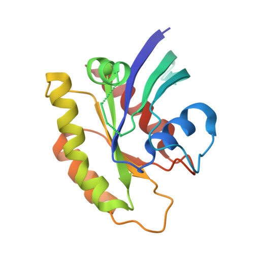
5UK9 - PubMed Abstract:
Structures of wild-type K-Ras from crystals obtained in the presence of guanosine triphosphate (GTP) or its analogs have remained elusive. Of the K-Ras mutants, only K-RasG12D and K-RasQ61H are available in the PDB representing the activated form of the GTPase not in complex with other proteins. We present the crystal structure of wild-type K-Ras bound to the GTP analog GppCH 2 p, with K-Ras in the state 1 conformation. Signatures of conformational states obtained by one-dimensional proton NMR confirm that K-Ras has a more substantial population of state 1 in solution than H-Ras, which predominantly favors state 2. The oncogenic mutant K-RasG12D favors state 2, changing the balance of conformational states in favor of interactions with effector proteins. Differences in the population of conformational states between K-Ras and H-Ras, as well as between K-Ras and its mutants, can provide a structural basis for focused targeting of the K-Ras isoform in cancer-specific strategies.
- Department of Chemistry & Chemical Biology, Northeastern University, Boston, MA 02115, USA.
Organizational Affiliation: