5UJO | pdb_00005ujo

X-Ray Crystal Structure of Ruthenocenyl-7-Aminodesacetoxycephalosporanic Acid Covalent Acyl-Enyzme Complex with CTX-M-14 E166A Beta-Lactamase

  • Classification: HYDROLASE
  • Organism(s): Escherichia coli
  • Expression System: Escherichia coli
  • Mutation(s): Yes 

  • Deposited: 2017-01-18 Released: 2017-02-08 
  • Deposition Author(s): Lewandowski, E.M., Chen, Y.
  • Funding Organization(s): National Institutes of Health/National Institute Of Allergy and Infectious Diseases (NIH/NIAID), Polish National Science Centre

Experimental Data Snapshot

  • Method: X-RAY DIFFRACTION
  • Resolution: 1.35 Å
  • R-Value Free: 
    0.150 (Depositor), 0.162 (DCC) 
  • R-Value Work: 
    0.125 (Depositor), 0.141 (DCC) 
  • R-Value Observed: 
    0.127 (Depositor) 

Starting Model: experimental
View more details

wwPDB Validation 3D Report Full Report

Validation slider image for 5UJO

Ligand Structure Quality Assessment 


This is version 1.6 of the entry. See complete history

Literature

Antibacterial Properties of Metallocenyl-7-ADCA Derivatives and Structure in Complex with CTX-Mbeta-Lactamase.

Lewandowski, E.M.Szczupak, L.Wong, S.Skiba, J.Guspiel, A.Solecka, J.Vrcek, V.Kowalski, K.Chen, Y.

(2017) Organometallics 36: 1673-1676

  • DOI: https://doi.org/10.1021/acs.organomet.6b00888
  • Primary Citation Related Structures: 
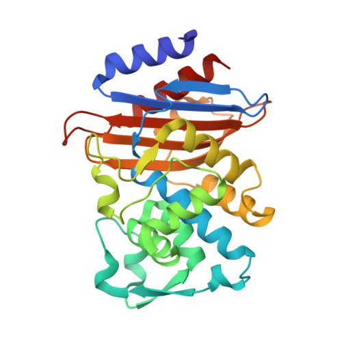
    5UJO

  • PubMed Abstract: 

    A series of six novel metallocenyl-7-ADCA (metallocenyl = ferrocenyl or ruthenocenyl; 7-ADCA = 7-aminodesacetoxycephalosporanic acid) conjugates were synthesized and their antibacterial properties evaluated by biochemical and microbiological assays. The ruthenocene derivatives showed a higher level of inhibition of DD-carboxypeptidase 64-575, a Penicillin Binding Protein (PBP), than the ferrocene derivatives and the reference compound penicillin G. Protein X-ray crystallographic analysis revealed a covalent acyl-enzyme complex of a ruthenocenyl compound with CTX-M β-lactamase E166A mutant, corresponding to a similar complex with PBPs responsible for the bactericidal activities of these compounds. Most interestingly, an intact compound was captured at the crystal-packing interface, elucidating for the first time the structure of a metallocenyl β-lactam compound that previously eluded small molecule crystallography. We propose that protein crystals, even from biologically unrelated molecules, can be utilized to determine structures of small molecules.


  • Organizational Affiliation
    • Department of Molecular Medicine, University of South Florida Morsani College of Medicine, 12901 Bruce B. Downs Blvd., Tampa, Florida 33612, United States.

Macromolecule Content 

  • Total Structure Weight: 58.09 kDa 
  • Atom Count: 5,029 
  • Modeled Residue Count: 522 
  • Deposited Residue Count: 526 
  • Unique protein chains: 1

Macromolecules

Find similar proteins by:|  3D Structure
Entity ID: 1
MoleculeChains  Sequence LengthOrganismDetailsImage
Beta-lactamase
A, B
263Escherichia coliMutation(s): 1 
Gene Names: blaCTX-MCTX-M-14
EC: 3.5.2.6
UniProt
Find proteins for Q9L5C8 (Escherichia coli)
Explore Q9L5C8 
Go to UniProtKB:  Q9L5C8
Entity Groups
Sequence Clusters30% Identity50% Identity70% Identity90% Identity95% Identity100% Identity
UniProt GroupQ9L5C8
Sequence Annotations
Expand
Reference Sequence

Small Molecules

Ligands 3 Unique
IDChains Name / Formula / InChI Key2D Diagram3D Interactions
8CY

Query on 8CY


Download:Ideal Coordinates CCD File
D [auth A],
G [auth B]
ruthenocenyl-7-aminodesacetoxycephalosporanic acid, bound form
C22 H15 N2 O6 Ru S
RODYUOCEXNRXSR-FZMMWMHASA-N
LSI

Query on LSI


Download:Ideal Coordinates CCD File
E [auth A],
H [auth B]
ruthenocenyl-7-aminodesacetoxycephalosporanic acid
C22 H22 N2 O5 Ru S
VVYXHQVDQWFSGH-DRUSRECESA-N
K

Query on K


Download:Ideal Coordinates CCD File
C [auth A],
F [auth B]
POTASSIUM ION
K
NPYPAHLBTDXSSS-UHFFFAOYSA-N

Experimental Data & Validation

Experimental Data

  • Method: X-RAY DIFFRACTION
  • Resolution: 1.35 Å
  • R-Value Free:  0.150 (Depositor), 0.162 (DCC) 
  • R-Value Work:  0.125 (Depositor), 0.141 (DCC) 
  • R-Value Observed: 0.127 (Depositor) 
Space Group: P 1 21 1
Unit Cell:
Length ( Å )Angle ( ˚ )
a = 44.961α = 90
b = 107.351β = 101.36
c = 47.956γ = 90
Software Package:
Software NamePurpose
REFMACrefinement
HKL-2000data reduction
SCALEPACKdata scaling
PHASERphasing

Structure Validation

View Full Validation Report


Ligand Structure Quality Assessment 


Entry History 

& Funding Information

Deposition Data


Funding OrganizationLocationGrant Number
National Institutes of Health/National Institute Of Allergy and Infectious Diseases (NIH/NIAID)United StatesAI03158
Polish National Science CentrePolandDEC-2013/11/B/ST5/00997

Revision History  (Full details and data files)

  • Version 1.0: 2017-02-08
    Type: Initial release
  • Version 1.1: 2017-07-19
    Changes: Database references
  • Version 1.2: 2017-09-13
    Changes: Author supporting evidence
  • Version 1.3: 2018-04-18
    Changes: Data collection, Database references
  • Version 1.4: 2019-12-11
    Changes: Author supporting evidence
  • Version 1.5: 2023-10-04
    Changes: Data collection, Database references, Derived calculations, Refinement description
  • Version 1.6: 2024-10-23
    Changes: Structure summary