5UHU | pdb_00005uhu

Solution conformation of cytochrome P450 MycG with mycinamicin IV bound


Experimental Data Snapshot

  • Method: SOLUTION NMR
  • Conformers Calculated: 1200 
  • Conformers Submitted: 
  • Selection Criteria: structures with the least restraint violations 

wwPDB Validation 3D Report Full Report

Validation slider image for 5UHU

This is version 1.3 of the entry. See complete history

Literature

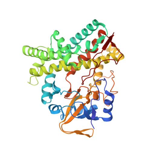
Solution Conformations and Dynamics of Substrate-Bound Cytochrome P450 MycG.

Tietz, D.R.Podust, L.M.Sherman, D.H.Pochapsky, T.C.

(2017) Biochemistry 56: 2701-2714

  • DOI: https://doi.org/10.1021/acs.biochem.7b00291
  • Primary Citation Related Structures: 
    5UHU

  • PubMed Abstract: 

    MycG is a P450 monooxygenase that catalyzes the sequential hydroxylation and epoxidation of mycinamicin IV (M-IV), the last two steps in the biosynthesis of mycinamicin II, a macrolide antibiotic isolated from Micromonospora griseorubida. The crystal structure of MycG with M-IV bound was previously determined but showed the bound substrate in an orientation that did not rationalize the observed regiochemistry of M-IV hydroxylation. Nuclear magnetic resonance paramagnetic relaxation enhancements provided evidence of an orientation of M-IV in the MycG active site more compatible with the observed chemistry, but substrate-induced changes in the enzyme structure were not characterized. We now describe the use of amide 1 H- 15 N residual dipolar couplings as experimental restraints in solvated "soft annealing" molecular dynamics simulations to generate solution structural ensembles of M-IV-bound MycG. Chemical shift perturbations, hydrogen-deuterium exchange, and 15 N relaxation behavior provide insight into the dynamic and electronic perturbations in the MycG structure in response to M-IV binding. The solution and crystallographic structures are compared, and the possibility that the crystallographic orientation of bound M-IV represents an inhibitory mode is discussed.


  • Organizational Affiliation
    • Skaggs School of Pharmacy and Pharmaceutical Sciences, University of California , San Diego, California 92093, United States.

Macromolecule Content 

  • Total Structure Weight: 45.7 kDa 
  • Atom Count: 3,187 
  • Modeled Residue Count: 393 
  • Deposited Residue Count: 397 
  • Unique protein chains: 1

Macromolecules

Find similar proteins by:|  3D Structure
Entity ID: 1
MoleculeChains  Sequence LengthOrganismDetailsImage
Mycinamicin IV hydroxylase/epoxidase397Micromonospora griseorubidaMutation(s): 0 
Gene Names: mycG
EC: 1.14
UniProt
Find proteins for Q59523 (Micromonospora griseorubida)
Explore Q59523 
Go to UniProtKB:  Q59523
Entity Groups
Sequence Clusters30% Identity50% Identity70% Identity90% Identity95% Identity100% Identity
UniProt GroupQ59523
Sequence Annotations
Expand
Reference Sequence

Small Molecules

Ligands 2 Unique
IDChains Name / Formula / InChI Key2D Diagram3D Interactions
MIV

Query on MIV


Download:Ideal Coordinates CCD File
C [auth A]MYCINAMICIN IV
C37 H61 N O11
DBTIHDIIXPQOFR-JMHKOBKLSA-N
HEM

Query on HEM


Download:Ideal Coordinates CCD File
B [auth A]PROTOPORPHYRIN IX CONTAINING FE
C34 H32 Fe N4 O4
KABFMIBPWCXCRK-RGGAHWMASA-L

Experimental Data & Validation

Experimental Data

  • Method: SOLUTION NMR
  • Conformers Calculated: 1200 
  • Conformers Submitted: 
  • Selection Criteria: structures with the least restraint violations 

Structure Validation

View Full Validation Report


Entry History 

& Funding Information

Deposition Data


Funding OrganizationLocationGrant Number
National Institutes of Health/National Institute of General Medical Sciences (NIH/NIGMS)United StatesR01GM44191

Revision History  (Full details and data files)

  • Version 1.0: 2017-08-23
    Type: Initial release
  • Version 1.1: 2017-09-27
    Changes: Author supporting evidence
  • Version 1.2: 2020-01-01
    Changes: Author supporting evidence, Data collection
  • Version 1.3: 2024-05-01
    Changes: Data collection, Database references