5UEW | pdb_00005uew

BRD2 Bromodomain2 with A-1360579


Experimental Data Snapshot

  • Method: X-RAY DIFFRACTION
  • Resolution: 1.83 Å
  • R-Value Free: 
    0.238 (Depositor), 0.250 (DCC) 
  • R-Value Work: 
    0.199 (Depositor), 0.210 (DCC) 
  • R-Value Observed: 
    0.201 (Depositor) 

wwPDB Validation 3D Report Full Report

Validation slider image for 5UEW

Ligand Structure Quality Assessment 


This is version 1.2 of the entry. See complete history

Literature

Fragment-Based, Structure-Enabled Discovery of Novel Pyridones and Pyridone Macrocycles as Potent Bromodomain and Extra-Terminal Domain (BET) Family Bromodomain Inhibitors.

Wang, L.Pratt, J.K.Soltwedel, T.Sheppard, G.S.Fidanze, S.D.Liu, D.Hasvold, L.A.Mantei, R.A.Holms, J.H.McClellan, W.J.Wendt, M.D.Wada, C.Frey, R.Hansen, T.M.Hubbard, R.Park, C.H.Li, L.Magoc, T.J.Albert, D.H.Lin, X.Warder, S.E.Kovar, P.Huang, X.Wilcox, D.Wang, R.Rajaraman, G.Petros, A.M.Hutchins, C.W.Panchal, S.C.Sun, C.Elmore, S.W.Shen, Y.Kati, W.M.McDaniel, K.F.

(2017) J Med Chem 60: 3828-3850

  • DOI: https://doi.org/10.1021/acs.jmedchem.7b00017
  • Primary Citation Related Structures: 
    5UEU, 5UEW, 5UEX, 5UEY, 5UEZ, 5UF0

  • PubMed Abstract: 

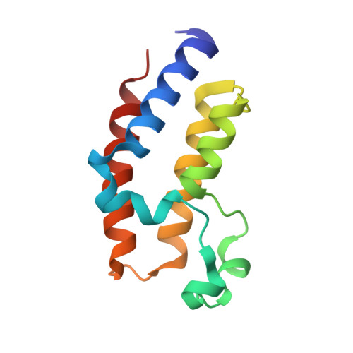
    Members of the BET family of bromodomain containing proteins have been identified as potential targets for blocking proliferation in a variety of cancer cell lines. A two-dimensional NMR fragment screen for binders to the bromodomains of BRD4 identified a phenylpyridazinone fragment with a weak binding affinity (1, K i = 160 μM). SAR investigation of fragment 1, aided by X-ray structure-based design, enabled the synthesis of potent pyridone and macrocyclic pyridone inhibitors exhibiting single digit nanomolar potency in both biochemical and cell based assays. Advanced analogs in these series exhibited high oral exposures in rodent PK studies and demonstrated significant tumor growth inhibition efficacy in mouse flank xenograft models.


  • Organizational Affiliation
    • AbbVie Inc. , 1 North Waukegan Road, North Chicago, Illinois 60064, United States.

Macromolecule Content 

  • Total Structure Weight: 26.85 kDa 
  • Atom Count: 2,021 
  • Modeled Residue Count: 222 
  • Deposited Residue Count: 222 
  • Unique protein chains: 1

Macromolecules

Find similar proteins by:|  3D Structure
Entity ID: 1
MoleculeChains  Sequence LengthOrganismDetailsImage
Bromodomain-containing protein 2
A, B
111Homo sapiensMutation(s): 0 
Gene Names: BRD2KIAA9001RING3
UniProt & NIH Common Fund Data Resources
Find proteins for P25440 (Homo sapiens)
Explore P25440 
Go to UniProtKB:  P25440
PHAROS:  P25440
GTEx:  ENSG00000204256 
Entity Groups
Sequence Clusters30% Identity50% Identity70% Identity90% Identity95% Identity100% Identity
UniProt GroupP25440
Sequence Annotations
Expand
Reference Sequence

Small Molecules

Ligands 1 Unique
IDChains Name / Formula / InChI Key2D Diagram3D Interactions
87D

Query on 87D


Download:Ideal Coordinates CCD File
C [auth A],
D [auth B]
N-[3-(4-methoxy-1-methyl-6-oxo-1,6-dihydropyridin-3-yl)-4-phenoxyphenyl]methanesulfonamide
C20 H20 N2 O5 S
RHNWTJMCFPHSQX-UHFFFAOYSA-N

Experimental Data & Validation

Experimental Data

  • Method: X-RAY DIFFRACTION
  • Resolution: 1.83 Å
  • R-Value Free:  0.238 (Depositor), 0.250 (DCC) 
  • R-Value Work:  0.199 (Depositor), 0.210 (DCC) 
  • R-Value Observed: 0.201 (Depositor) 
Space Group: P 1
Unit Cell:
Length ( Å )Angle ( ˚ )
a = 35.996α = 67.4
b = 41.601β = 66.9
c = 45.756γ = 74.85
Software Package:
Software NamePurpose
XDSdata reduction
SCALAdata scaling
Cootmodel building
BUSTERrefinement

Structure Validation

View Full Validation Report


Ligand Structure Quality Assessment 


Entry History 

Deposition Data

  • Released Date: 2017-05-10 
  • Deposition Author(s): Park, C.H.

Revision History  (Full details and data files)

  • Version 1.0: 2017-05-10
    Type: Initial release
  • Version 1.1: 2017-05-24
    Changes: Database references
  • Version 1.2: 2024-03-06
    Changes: Data collection, Database references