5UC6

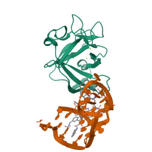
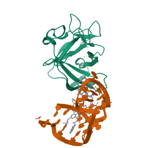
Structural insights into IL-1 alpha recognition by a naphthyl-modified aptamer that mimics IL-1RI Domain III


Experimental Data Snapshot

  • Method: X-RAY DIFFRACTION
  • Resolution: 2.10 Å
  • R-Value Free: 0.219 
  • R-Value Work: 0.178 
  • R-Value Observed: 0.179 

Starting Model: experimental
View more details

wwPDB Validation   3D Report Full Report


This is version 3.2 of the entry. See complete history


Literature

Structural basis for IL-1 alpha recognition by a modified DNA aptamer that specifically inhibits IL-1 alpha signaling.

Ren, X.Gelinas, A.D.von Carlowitz, I.Janjic, N.Pyle, A.M.

(2017) Nat Commun 8: 810-810

  • DOI: https://doi.org/10.1038/s41467-017-00864-2
  • Primary Citation of Related Structures:  
    5UC6

  • PubMed Abstract: 

    IL-1α is an essential cytokine that contributes to inflammatory responses and is implicated in various forms of pathogenesis and cancer. Here we report a naphthyl modified DNA aptamer that specifically binds IL-1α and inhibits its signaling pathway. By solving the crystal structure of the IL-1α/aptamer, we provide a high-resolution structure of this critical cytokine and we reveal its functional interaction interface with high-affinity ligands. The non-helical aptamer, which represents a highly compact nucleic acid structure, contains a wealth of new conformational features, including an unknown form of G-quadruplex. The IL-1α/aptamer interface is composed of unusual polar and hydrophobic elements, along with an elaborate hydrogen bonding network that is mediated by sodium ion. IL-1α uses the same interface to interact with both the aptamer and its cognate receptor IL-1RI, thereby suggesting a novel route to immunomodulatory therapeutics.The cytokine interleukin 1α (IL-1α) plays an important role in inflammatory processes. Here the authors use SELEX to generate a modified DNA aptamer which specifically binds IL-1α, present the structure of the IL-1α/aptamer complex and show that this aptamer inhibits the IL-1α signaling pathway.


  • Organizational Affiliation

    Department of Molecular, Cellular, and Developmental Biology, Yale University, 219 Prospect Street, New Haven, CT, 06511, USA.


Macromolecules

Find similar proteins by:  (by identity cutoff)  |  3D Structure
Entity ID: 1
MoleculeChains Sequence LengthOrganismDetailsImage
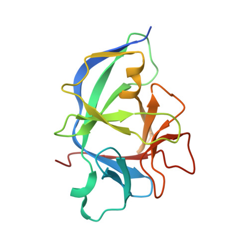
Interleukin-1 alpha159Homo sapiensMutation(s): 0 
Gene Names: IL1AIL1F1
UniProt & NIH Common Fund Data Resources
Find proteins for P01583 (Homo sapiens)
Explore P01583 
Go to UniProtKB:  P01583
PHAROS:  P01583
GTEx:  ENSG00000115008 
Entity Groups  
Sequence Clusters30% Identity50% Identity70% Identity90% Identity95% Identity100% Identity
UniProt GroupP01583
Sequence Annotations
Expand
  • Reference Sequence

Find similar nucleic acids by:  Sequence   |   3D Structure  

Entity ID: 2
MoleculeChains LengthOrganismImage
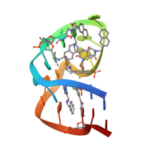
DNA (5'-D(*CP*G)-R(P*(85Y))-D(P*GP*AP*G)-R(P*(85Y)P*(85Y))-D(P*A)-R(P*(85Y))-D(P*GP*GP*G)-R(P*(85Y)P*(85Y))-D(P*AP*GP*AP*G)-R(P*(85Y))-D(P*CP*GP*(ATD))-3')23synthetic construct
Sequence Annotations
Expand
  • Reference Sequence
Experimental Data & Validation

Experimental Data

  • Method: X-RAY DIFFRACTION
  • Resolution: 2.10 Å
  • R-Value Free: 0.219 
  • R-Value Work: 0.178 
  • R-Value Observed: 0.179 
  • Space Group: P 31 2 1
Unit Cell:
Length ( Å )Angle ( ˚ )
a = 74.79α = 90
b = 74.79β = 90
c = 86.36γ = 120
Software Package:
Software NamePurpose
PHENIXrefinement
iMOSFLMdata reduction
SCALAdata scaling
PHASERphasing

Structure Validation

View Full Validation Report


Entry History & Funding Information

Deposition Data


Funding OrganizationLocationGrant Number
Howard Hughes Medical Institute (HHMI)United States--

Revision History  (Full details and data files)

  • Version 1.0: 2017-10-11
    Type: Initial release
  • Version 1.1: 2017-10-18
    Changes: Database references
  • Version 2.0: 2017-11-15
    Changes: Atomic model, Derived calculations, Non-polymer description, Structure summary
  • Version 3.0: 2018-08-22
    Changes: Atomic model, Data collection, Derived calculations
  • Version 3.1: 2019-11-20
    Changes: Author supporting evidence
  • Version 3.2: 2023-10-04
    Changes: Data collection, Database references, Derived calculations, Refinement description