5UBX | pdb_00005ubx

Crystal structure of a mutant mIgG2b Fc heterodimer in complex with Protein A peptide analog Z34C


Experimental Data Snapshot

  • Method: X-RAY DIFFRACTION
  • Resolution: 2.70 Å
  • R-Value Free: 
    0.257 (Depositor), 0.258 (DCC) 
  • R-Value Work: 
    0.209 (Depositor), 0.222 (DCC) 
  • R-Value Observed: 
    0.212 (Depositor) 

Starting Model: experimental
View more details

wwPDB Validation 3D Report Full Report

Validation slider image for 5UBX

This is version 2.2 of the entry. See complete history

Literature

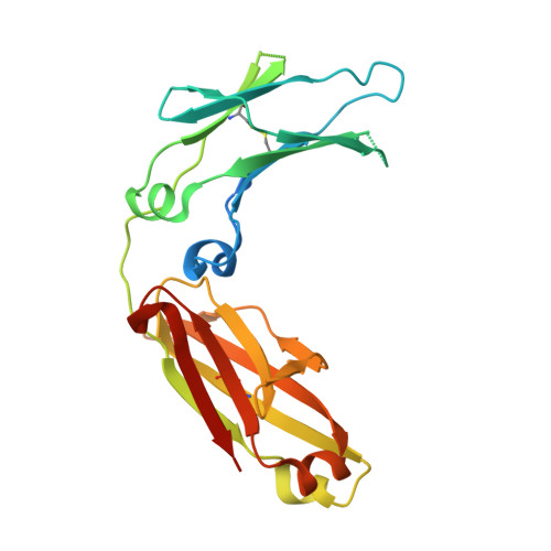
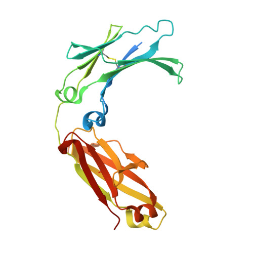
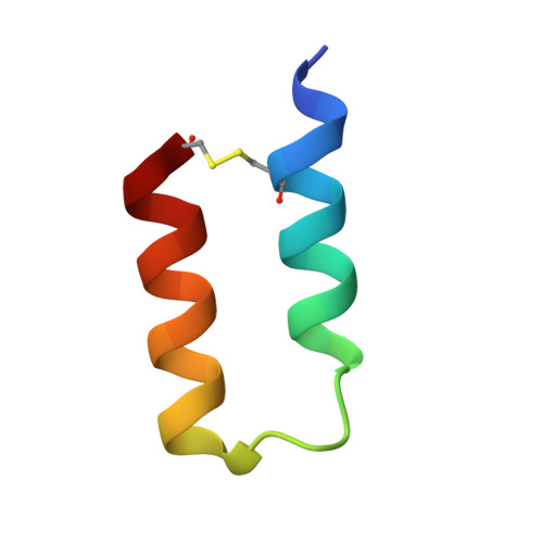
Modulation of protein A binding allows single-step purification of mouse bispecific antibodies that retain FcRn binding.

Zwolak, A.Armstrong, A.A.Tam, S.H.Pardinas, J.R.Goulet, D.R.Zheng, S.Brosnan, K.Emmell, E.Luo, J.Gilliland, G.L.Chiu, M.L.

(2017) MAbs 9: 1306-1316

  • DOI: https://doi.org/10.1080/19420862.2017.1375639
  • Primary Citation Related Structures: 
    5UBX

  • PubMed Abstract: 

    The increased number of bispecific antibodies (BsAb) under therapeutic development has resulted in a need for mouse surrogate BsAbs. Here, we describe a one-step method for generating highly pure mouse BsAbs suitable for in vitro and in vivo studies. We identify two mutations in the mouse IgG2a and IgG2b Fc region: one that eliminates protein A binding and one that enhances protein A binding by 8-fold. We show that BsAbs harboring these mutations can be purified from the residual parental monoclonal antibodies in one step using protein A affinity chromatography. The structural basis for the effects of these mutations was analyzed by X-ray crystallography. While the mutation that disrupted protein A binding also inhibited FcRn interaction, a bispecific mutant in which one subunit retained the ability to bind protein A could still interact with FcRn. Pharmacokinetic analysis of the serum half-lives of the mutants showed that the mutant BsAb had a serum half-life comparable to a wild-type Ab. The results describe a rapid method for generating panels of mouse BsAbs that could be used in mouse studies.


  • Organizational Affiliation
    • a Biologics Discovery , Janssen Research & Development, LLC , Spring House , PA , USA.

Macromolecule Content 

  • Total Structure Weight: 59.28 kDa 
  • Atom Count: 3,453 
  • Modeled Residue Count: 426 
  • Deposited Residue Count: 500 
  • Unique protein chains: 3

Macromolecules

Find similar proteins by:|  3D Structure
Entity ID: 1
MoleculeChains  Sequence LengthOrganismDetailsImage
Ig gamma-2B chain C region230Mus musculusMutation(s): 4 
Gene Names: Igh-3
UniProt
Find proteins for P01867 (Mus musculus)
Explore P01867 
Go to UniProtKB:  P01867
Entity Groups
Sequence Clusters30% Identity50% Identity70% Identity90% Identity95% Identity100% Identity
UniProt GroupP01867
Sequence Annotations
Expand
Reference Sequence
Find similar proteins by:|  3D Structure
Entity ID: 2
MoleculeChains  Sequence LengthOrganismDetailsImage
Ig gamma-2B chain C region236Mus musculusMutation(s): 2 
Gene Names: Igh-3
UniProt
Find proteins for P01867 (Mus musculus)
Explore P01867 
Go to UniProtKB:  P01867
Entity Groups
Sequence Clusters30% Identity50% Identity70% Identity90% Identity95% Identity100% Identity
UniProt GroupP01867
Glycosylation
Glycosylation Sites: 1Go to GlyGen: P01867-1
Sequence Annotations
Expand
Reference Sequence
Find similar proteins by:|  3D Structure
Entity ID: 3
MoleculeChains  Sequence LengthOrganismDetailsImage
Peptide analog of B-domain from Protein A - Z34CC [auth Z]34Staphylococcus aureusMutation(s): 0 

Oligosaccharides

Help  
Entity ID: 4
MoleculeChains Length2D Diagram GlycosylationD Interactions
2-acetamido-2-deoxy-beta-D-glucopyranose-(1-2)-alpha-D-mannopyranose-(1-3)-[2-acetamido-2-deoxy-beta-D-glucopyranose-(1-2)-alpha-D-mannopyranose-(1-6)]beta-D-mannopyranose-(1-4)-2-acetamido-2-deoxy-beta-D-glucopyranoseD [auth C]6N/A
Glycosylation Resources
GlyTouCan: G55061AW
GlyCosmos: G55061AW
GlyGen: G55061AW
Entity ID: 5
MoleculeChains Length2D Diagram GlycosylationD Interactions
2-acetamido-2-deoxy-beta-D-glucopyranose-(1-2)-alpha-D-mannopyranose-(1-3)-[2-acetamido-2-deoxy-beta-D-glucopyranose-(1-2)-alpha-D-mannopyranose-(1-6)]beta-D-mannopyranose-(1-4)-2-acetamido-2-deoxy-beta-D-glucopyranose-(1-4)-[alpha-L-fucopyranose-(1-6)]2-acetamido-2-deoxy-beta-D-glucopyranoseE [auth D]8N-Glycosylation
Glycosylation Resources
GlyTouCan: G80858MF
GlyCosmos: G80858MF
GlyGen: G80858MF

Experimental Data & Validation

Experimental Data

  • Method: X-RAY DIFFRACTION
  • Resolution: 2.70 Å
  • R-Value Free:  0.257 (Depositor), 0.258 (DCC) 
  • R-Value Work:  0.209 (Depositor), 0.222 (DCC) 
  • R-Value Observed: 0.212 (Depositor) 
Space Group: P 43 21 2
Unit Cell:
Length ( Å )Angle ( ˚ )
a = 101.49α = 90
b = 101.49β = 90
c = 135.07γ = 90
Software Package:
Software NamePurpose
PHENIXrefinement
XDSdata reduction
XDSdata scaling
PHASERphasing

Structure Validation

View Full Validation Report


Entry History 

Deposition Data

Revision History  (Full details and data files)

  • Version 1.0: 2017-09-20
    Type: Initial release
  • Version 1.1: 2017-10-04
    Changes: Database references
  • Version 1.2: 2017-11-08
    Changes: Database references
  • Version 2.0: 2020-07-29
    Type: Remediation
    Reason: Carbohydrate remediation
    Changes: Advisory, Atomic model, Data collection, Derived calculations, Structure summary
  • Version 2.1: 2023-10-04
    Changes: Data collection, Database references, Derived calculations, Refinement description, Structure summary
  • Version 2.2: 2024-11-20
    Changes: Structure summary