5UBQ | pdb_00005ubq

Cryo-EM structure of ciliary microtubule doublet


Experimental Data Snapshot

  • Method: ELECTRON MICROSCOPY
  • Resolution: 5.70 Å
  • Aggregation State: FILAMENT 
  • Reconstruction Method: SINGLE PARTICLE 

wwPDB Validation 3D Report Full Report

Validation slider image for 5UBQ

This is version 1.6 of the entry. See complete history

Literature

Subnanometre-resolution structure of the doublet microtubule reveals new classes of microtubule-associated proteins.

Ichikawa, M.Liu, D.Kastritis, P.L.Basu, K.Hsu, T.C.Yang, S.Bui, K.H.

(2017) Nat Commun 8: 15035-15035

  • DOI: https://doi.org/10.1038/ncomms15035
  • Primary Citation Related Structures: 
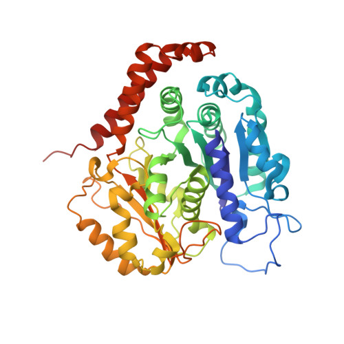
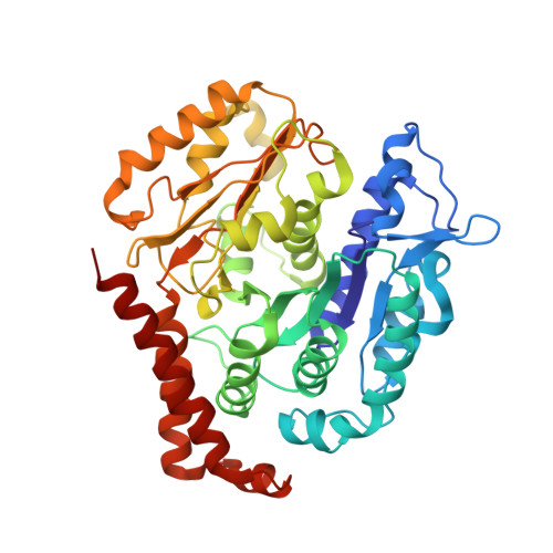
    5UBQ, 5UCY

  • PubMed Abstract: 

    Cilia are ubiquitous, hair-like appendages found in eukaryotic cells that carry out functions of cell motility and sensory reception. Cilia contain an intriguing cytoskeletal structure, termed the axoneme that consists of nine doublet microtubules radially interlinked and longitudinally organized in multiple specific repeat units. Little is known, however, about how the axoneme allows cilia to be both actively bendable and sturdy or how it is assembled. To answer these questions, we used cryo-electron microscopy to structurally analyse several of the repeating units of the doublet at sub-nanometre resolution. This structural detail enables us to unambiguously assign α- and β-tubulins in the doublet microtubule lattice. Our study demonstrates the existence of an inner sheath composed of different kinds of microtubule inner proteins inside the doublet that likely stabilizes the structure and facilitates the specific building of the B-tubule.


  • Organizational Affiliation
    • Department of Anatomy and Cell Biology, McGill University, Montréal, Québec, Canada H3A 0C7.

Macromolecule Content 

  • Total Structure Weight: 293.36 kDa 
  • Atom Count: 20,538 
  • Modeled Residue Count: 2,610 
  • Deposited Residue Count: 2,610 
  • Unique protein chains: 2

Macromolecules

Find similar proteins by:|  3D Structure
Entity ID: 1
MoleculeChains  Sequence LengthOrganismDetailsImage
Tubulin alpha chainA [auth C],
C [auth E],
E [auth A]
441Tetrahymena thermophilaMutation(s): 0 
EC: 3.6.5
UniProt
Find proteins for P41351 (Tetrahymena thermophila)
Explore P41351 
Go to UniProtKB:  P41351
Entity Groups
Sequence Clusters30% Identity50% Identity70% Identity90% Identity95% Identity100% Identity
UniProt GroupP41351
Sequence Annotations
Expand
Reference Sequence
Find similar proteins by:|  3D Structure
Entity ID: 2
MoleculeChains  Sequence LengthOrganismDetailsImage
Tubulin beta chainB [auth D],
D [auth F],
F [auth B]
429Tetrahymena thermophilaMutation(s): 0 
UniProt
Find proteins for P41352 (Tetrahymena thermophila)
Explore P41352 
Go to UniProtKB:  P41352
Entity Groups
Sequence Clusters30% Identity50% Identity70% Identity90% Identity95% Identity100% Identity
UniProt GroupP41352
Sequence Annotations
Expand
Reference Sequence

Small Molecules

Ligands 3 Unique
IDChains Name / Formula / InChI Key2D Diagram3D Interactions
GTP

Query on GTP


Download:Ideal Coordinates CCD File
G [auth C],
J [auth E],
M [auth A]
GUANOSINE-5'-TRIPHOSPHATE
C10 H16 N5 O14 P3
XKMLYUALXHKNFT-UUOKFMHZSA-N
GDP

Query on GDP


Download:Ideal Coordinates CCD File
I [auth D],
L [auth F],
O [auth B]
GUANOSINE-5'-DIPHOSPHATE
C10 H15 N5 O11 P2
QGWNDRXFNXRZMB-UUOKFMHZSA-N
MG

Query on MG


Download:Ideal Coordinates CCD File
H [auth C],
K [auth E],
N [auth A]
MAGNESIUM ION
Mg
JLVVSXFLKOJNIY-UHFFFAOYSA-N

Experimental Data & Validation

Experimental Data

  • Method: ELECTRON MICROSCOPY
  • Resolution: 5.70 Å
  • Aggregation State: FILAMENT 
  • Reconstruction Method: SINGLE PARTICLE 
EM Software:
TaskSoftware PackageVersion
RECONSTRUCTIONRELION1.4,2.0
MODEL REFINEMENTPHENIX

Structure Validation

View Full Validation Report


Entry History 

& Funding Information

Deposition Data


Funding OrganizationLocationGrant Number
Natural Sciences and Engineering Research Council (NSERC, Canada)Canada69462

Revision History  (Full details and data files)

  • Version 1.0: 2017-05-10
    Type: Initial release
  • Version 1.1: 2017-05-17
    Changes: Database references
  • Version 1.2: 2017-09-27
    Changes: Author supporting evidence, Data collection
  • Version 1.3: 2018-05-16
    Changes: Data collection, Derived calculations
  • Version 1.4: 2018-07-18
    Changes: Data collection
  • Version 1.5: 2020-01-15
    Changes: Author supporting evidence
  • Version 1.6: 2024-12-25
    Changes: Advisory, Data collection, Database references, Derived calculations, Structure summary