5U8Z | pdb_00005u8z

Structure of Fe-CAO1 in complex with beta-fluororesveratrol

  • Classification: OXIDOREDUCTASE
  • Organism(s): Neurospora crassa OR74A
  • Expression System: Escherichia coli
  • Mutation(s): No 

  • Deposited: 2016-12-15 Released: 2017-05-31 
  • Deposition Author(s): Sui, X., Palczewski, K., Kiser, P.D.
  • Funding Organization(s): National Institutes of Health/National Eye Institute (NIH/NEI), National Institutes of Health/National Cancer Institute (NIH/NCI), National Institutes of Health/National Institute of General Medical Sciences (NIH/NIGMS), Burroughs Wellcome Fund, Dept of Veterans Affairs

Experimental Data Snapshot

  • Method: X-RAY DIFFRACTION
  • Resolution: 1.90 Å
  • R-Value Free: 
    0.213 (Depositor), 0.222 (DCC) 
  • R-Value Work: 
    0.182 (Depositor), 0.191 (DCC) 
  • R-Value Observed: 
    0.183 (Depositor) 

Starting Model: experimental
View more details

wwPDB Validation 3D Report Full Report

Validation slider image for 5U8Z

Ligand Structure Quality Assessment 


This is version 1.5 of the entry. See complete history

Literature

Structure and Spectroscopy of Alkene-Cleaving Dioxygenases Containing an Atypically Coordinated Non-Heme Iron Center.

Sui, X.Weitz, A.C.Farquhar, E.R.Badiee, M.Banerjee, S.von Lintig, J.Tochtrop, G.P.Palczewski, K.Hendrich, M.P.Kiser, P.D.

(2017) Biochemistry 56: 2836-2852

  • DOI: https://doi.org/10.1021/acs.biochem.7b00251
  • Primary Citation Related Structures: 
    5U8X, 5U8Y, 5U8Z, 5U90, 5U97

  • PubMed Abstract: 

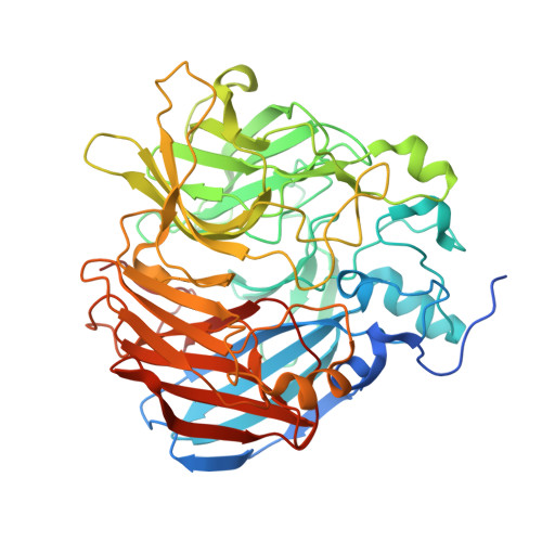
    Carotenoid cleavage oxygenases (CCOs) are non-heme iron enzymes that catalyze scission of alkene groups in carotenoids and stilbenoids to form biologically important products. CCOs possess a rare four-His iron center whose resting-state structure and interaction with substrates are incompletely understood. Here, we address this knowledge gap through a comprehensive structural and spectroscopic study of three phyletically diverse CCOs. The crystal structure of a fungal stilbenoid-cleaving CCO, CAO1, reveals strong similarity between its iron center and those of carotenoid-cleaving CCOs, but with a markedly different substrate-binding cleft. These enzymes all possess a five-coordinate high-spin Fe(II) center with resting-state Fe-His bond lengths of ∼2.15 Å. This ligand set generates an iron environment more electropositive than those of other non-heme iron dioxygenases as observed by Mössbauer isomer shifts. Dioxygen (O 2 ) does not coordinate iron in the absence of substrate. Substrates bind away (∼4.7 Å) from the iron and have little impact on its electronic structure, thus excluding coordination-triggered O 2 binding. However, substrate binding does perturb the spectral properties of CCO Fe-NO derivatives, indicating proximate organic substrate and O 2 -binding sites, which might influence Fe-O 2 interactions. Together, these data provide a robust description of the CCO iron center and its interactions with substrates and substrate mimetics that illuminates commonalities as well as subtle and profound structural differences within the CCO family.


  • Organizational Affiliation
    • Department of Pharmacology, School of Medicine, Case Western Reserve University , 10900 Euclid Avenue, Cleveland, Ohio 44106, United States.

Macromolecule Content 

  • Total Structure Weight: 239.5 kDa 
  • Atom Count: 17,697 
  • Modeled Residue Count: 1,991 
  • Deposited Residue Count: 2,104 
  • Unique protein chains: 1

Macromolecules

Find similar proteins by:|  3D Structure
Entity ID: 1
MoleculeChains  Sequence LengthOrganismDetailsImage
Carotenoid oxygenase 1
A, B, C, D
526Neurospora crassa OR74AMutation(s): 0 
Gene Names: cao-1NCU07008
EC: 1.13.11
UniProt
Find proteins for Q7S860 (Neurospora crassa (strain ATCC 24698 / 74-OR23-1A / CBS 708.71 / DSM 1257 / FGSC 987))
Explore Q7S860 
Go to UniProtKB:  Q7S860
Entity Groups
Sequence Clusters30% Identity50% Identity70% Identity90% Identity95% Identity100% Identity
UniProt GroupQ7S860
Sequence Annotations
Expand
Reference Sequence

Small Molecules

Ligands 4 Unique
IDChains Name / Formula / InChI Key2D Diagram3D Interactions
83D

Query on 83D


Download:Ideal Coordinates CCD File
F [auth A],
J [auth B],
M [auth C],
P [auth D]
5-[(Z)-2-fluoro-2-(4-hydroxyphenyl)ethenyl]benzene-1,3-diol
C14 H11 F O3
PNBPFKSSWYNPPD-AUWJEWJLSA-N
DMS

Query on DMS


Download:Ideal Coordinates CCD File
H [auth A],
R [auth D]
DIMETHYL SULFOXIDE
C2 H6 O S
IAZDPXIOMUYVGZ-UHFFFAOYSA-N
FE2

Query on FE2


Download:Ideal Coordinates CCD File
E [auth A],
I [auth B],
L [auth C],
O [auth D]
FE (II) ION
Fe
CWYNVVGOOAEACU-UHFFFAOYSA-N
CL

Query on CL


Download:Ideal Coordinates CCD File
G [auth A],
K [auth B],
N [auth C],
Q [auth D]
CHLORIDE ION
Cl
VEXZGXHMUGYJMC-UHFFFAOYSA-M

Experimental Data & Validation

Experimental Data

  • Method: X-RAY DIFFRACTION
  • Resolution: 1.90 Å
  • R-Value Free:  0.213 (Depositor), 0.222 (DCC) 
  • R-Value Work:  0.182 (Depositor), 0.191 (DCC) 
  • R-Value Observed: 0.183 (Depositor) 
Space Group: P 32 2 1
Unit Cell:
Length ( Å )Angle ( ˚ )
a = 101.037α = 90
b = 101.037β = 90
c = 448.622γ = 120
Software Package:
Software NamePurpose
REFMACrefinement
XDSdata reduction
XDSdata scaling

Structure Validation

View Full Validation Report


Ligand Structure Quality Assessment 


Entry History 

& Funding Information

Deposition Data


Funding OrganizationLocationGrant Number
National Institutes of Health/National Eye Institute (NIH/NEI)United StatesEY009339
National Institutes of Health/National Eye Institute (NIH/NEI)United StatesEY020551
National Institutes of Health/National Cancer Institute (NIH/NCI)CA157735
National Institutes of Health/National Institute of General Medical Sciences (NIH/NIGMS)GM77387
Burroughs Wellcome FundUnited States1015187
Dept of Veterans AffairsUnited StatesBX002683

Revision History  (Full details and data files)

  • Version 1.0: 2017-05-31
    Type: Initial release
  • Version 1.1: 2017-06-21
    Changes: Database references, Structure summary
  • Version 1.2: 2017-09-13
    Changes: Author supporting evidence
  • Version 1.3: 2019-12-11
    Changes: Author supporting evidence
  • Version 1.4: 2022-04-13
    Changes: Author supporting evidence, Database references, Derived calculations
  • Version 1.5: 2023-10-04
    Changes: Data collection, Refinement description