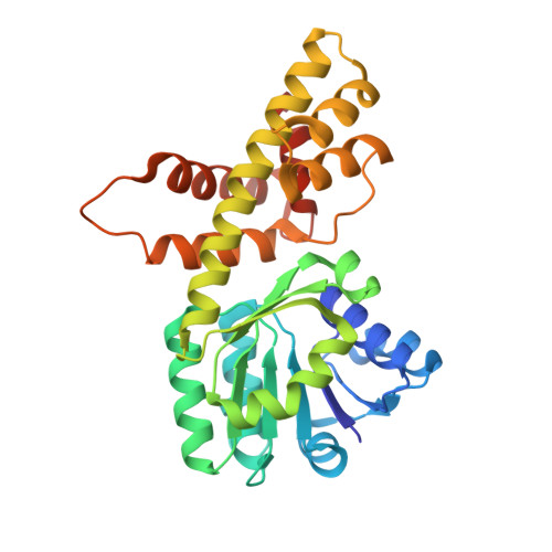
5U5G

Psf3 in complex with NADP+ and 2-OPP


Experimental Data Snapshot

  • Method: X-RAY DIFFRACTION
  • Resolution: 2.05 Å
  • R-Value Free: 0.207 
  • R-Value Work: 0.170 
  • R-Value Observed: 0.172 

wwPDB Validation   3D Report Full Report


Ligand Structure Quality Assessment 


This is version 1.5 of the entry. See complete history


Literature

Characterization of Two Late-Stage Enzymes Involved in Fosfomycin Biosynthesis in Pseudomonads.

Olivares, P.Ulrich, E.C.Chekan, J.R.van der Donk, W.A.Nair, S.K.

(2017) ACS Chem Biol 12: 456-463

  • DOI: https://doi.org/10.1021/acschembio.6b00939
  • Primary Citation of Related Structures:  
    5U55, 5U57, 5U58, 5U5D, 5U5G

  • PubMed Abstract: 

    The broad-spectrum phosphonate antibiotic fosfomycin is currently in use for clinical treatment of infections caused by both Gram-positive and Gram-negative uropathogens. The antibiotic is biosynthesized by various streptomycetes, as well as by pseudomonads. Notably, the biosynthetic strategies used by the two genera share only two steps: the first step in which primary metabolite phosphoenolpyruvate (PEP) is converted to phosphonopyruvate (PnPy) and the terminal step in which 2-hydroxypropylphosphonate (2-HPP) is converted to fosfomycin. Otherwise, distinct enzymatic paths are employed. Here, we biochemically confirm the last two steps in the fosfomycin biosynthetic pathway of Pseudomonas syringae PB-5123, showing that Psf3 performs the reduction of 2-oxopropylphosphonate (2-OPP) to (S)-2-HPP, followed by the Psf4-catalyzed epoxidation of (S)-2-HPP to fosfomycin. Psf4 can also accept (R)-2-HPP as a substrate but instead performs an oxidation to make 2-OPP. We show that the combined activities of Psf3 and Psf4 can be used to convert racemic 2-HPP to fosfomycin in an enantioconvergent process. X-ray structures of each enzyme with bound substrates provide insights into the stereospecificity of each conversion. These studies shed light on the reaction mechanisms of the two terminal enzymes in a distinct pathway employed by pseudomonads for the production of a potent antimicrobial agent.


  • Organizational Affiliation

    Department of Biochemistry, ‡Department of Chemistry, §Carl R. Woese Institute for Genomic Biology, ∥Howard Hughes Medical Institute, and ⊥Center for Biophysics and Computational Biology, University of Illinois at Urbana-Champaign , Urbana, Illinois 61801, United States.


Macromolecules
Find similar proteins by:  (by identity cutoff)  |  3D Structure
Entity ID: 1
MoleculeChains Sequence LengthOrganismDetailsImage
6-phosphogluconate dehydrogenase
A, B, C, D
295Pseudomonas syringaeMutation(s): 0 
UniProt
Find proteins for I6R485 (Pseudomonas syringae)
Explore I6R485 
Go to UniProtKB:  I6R485
Entity Groups  
Sequence Clusters30% Identity50% Identity70% Identity90% Identity95% Identity100% Identity
UniProt GroupI6R485
Sequence Annotations
Expand
  • Reference Sequence
Small Molecules
Ligands 3 Unique
IDChains Name / Formula / InChI Key2D Diagram3D Interactions
NAP
Query on NAP

Download Ideal Coordinates CCD File 
E [auth A],
H [auth B],
J [auth C],
M [auth D]
NADP NICOTINAMIDE-ADENINE-DINUCLEOTIDE PHOSPHATE
C21 H28 N7 O17 P3
XJLXINKUBYWONI-NNYOXOHSSA-N
PGE
Query on PGE

Download Ideal Coordinates CCD File 
G [auth A],
L [auth C]
TRIETHYLENE GLYCOL
C6 H14 O4
ZIBGPFATKBEMQZ-UHFFFAOYSA-N
7VD
Query on 7VD

Download Ideal Coordinates CCD File 
F [auth A],
I [auth B],
K [auth C],
N [auth D]
(2-oxopropyl)phosphonic acid
C3 H7 O4 P
DVSHXLWNRCEDCW-UHFFFAOYSA-N
Experimental Data & Validation

Experimental Data

  • Method: X-RAY DIFFRACTION
  • Resolution: 2.05 Å
  • R-Value Free: 0.207 
  • R-Value Work: 0.170 
  • R-Value Observed: 0.172 
  • Space Group: P 1 21 1
Unit Cell:
Length ( Å )Angle ( ˚ )
a = 46.076α = 90
b = 87.369β = 92.04
c = 164.011γ = 90
Software Package:
Software NamePurpose
PHENIXrefinement
HKL-2000data reduction
HKL-2000data scaling
PHASERphasing

Structure Validation

View Full Validation Report


Ligand Structure Quality Assessment 


Entry History & Funding Information

Deposition Data


Funding OrganizationLocationGrant Number
National Institutes of Health/National Institute of General Medical Sciences (NIH/NIGMS)United StatesPO1 GM077596

Revision History  (Full details and data files)

  • Version 1.0: 2017-01-04
    Type: Initial release
  • Version 1.1: 2017-03-01
    Changes: Database references
  • Version 1.2: 2017-09-20
    Changes: Author supporting evidence
  • Version 1.3: 2017-11-01
    Changes: Author supporting evidence
  • Version 1.4: 2020-01-01
    Changes: Author supporting evidence
  • Version 1.5: 2024-03-06
    Changes: Data collection, Database references