5U2P | pdb_00005u2p

The crystal structure of Tp0737 from Treponema pallidum


Experimental Data Snapshot

  • Method: X-RAY DIFFRACTION
  • Resolution: 1.76 Å
  • R-Value Free: 
    0.222 (Depositor), 0.224 (DCC) 
  • R-Value Work: 
    0.181 (Depositor), 0.183 (DCC) 
  • R-Value Observed: 
    0.183 (Depositor) 

wwPDB Validation 3D Report Full Report

Validation slider image for 5U2P

This is version 1.4 of the entry. See complete history

Literature

Functional clues from the crystal structure of an orphan periplasmic ligand-binding protein from Treponema pallidum.

Brautigam, C.A.Deka, R.K.Liu, W.Z.Tomchick, D.R.Norgard, M.V.

(2017) Protein Sci 26: 847-856

  • DOI: https://doi.org/10.1002/pro.3133
  • Primary Citation Related Structures: 
    5U2P

  • PubMed Abstract: 

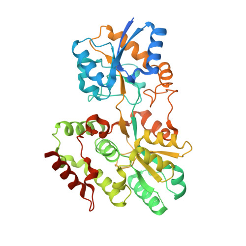
    The spirochete Treponema pallidum is the causative agent of syphilis, a sexually transmitted infection of major global importance. Other closely related subspecies of Treponema also are the etiological agents of the endemic treponematoses, such as yaws, pinta, and bejel. The inability of T. pallidum and its close relatives to be cultured in vitro has prompted efforts to characterize T. pallidum's proteins structurally and biophysically, particularly those potentially relevant to treponemal membrane biology, with the goal of possibly revealing the functions of those proteins. This report describes the structure of the treponemal protein Tp0737; this polypeptide has a fold characteristic of a class of periplasmic ligand-binding proteins associated with ABC-type transporters. Although no ligand for the protein was observed in electron-density maps, and thus the nature of the native ligand remains obscure, the structural data described herein provide a foundation for further efforts to elucidate the ligand and thus the function of this protein in T. pallidum.


  • Organizational Affiliation
    • Department of Biophysics, The University of Texas Southwestern Medical Center, Dallas, Texas, 75390.

Macromolecule Content 

  • Total Structure Weight: 47.93 kDa 
  • Atom Count: 3,211 
  • Modeled Residue Count: 381 
  • Deposited Residue Count: 428 
  • Unique protein chains: 1

Macromolecules

Find similar proteins by:|  3D Structure
Entity ID: 1
MoleculeChains  Sequence LengthOrganismDetailsImage
Sugar ABC transporter substrate-binding protein428Treponema pallidum subsp. pallidumMutation(s): 0 
Gene Names: A8P32_03680

Experimental Data & Validation

Experimental Data

  • Method: X-RAY DIFFRACTION
  • Resolution: 1.76 Å
  • R-Value Free:  0.222 (Depositor), 0.224 (DCC) 
  • R-Value Work:  0.181 (Depositor), 0.183 (DCC) 
  • R-Value Observed: 0.183 (Depositor) 
Space Group: P 31 2 1
Unit Cell:
Length ( Å )Angle ( ˚ )
a = 55.925α = 90
b = 55.925β = 90
c = 237.632γ = 120
Software Package:
Software NamePurpose
PHENIXrefinement
HKL-2000data reduction
HKL-2000data scaling
MLPHAREphasing

Structure Validation

View Full Validation Report


Entry History 

& Funding Information

Deposition Data


Funding OrganizationLocationGrant Number
National Institutes of Health/National Institute Of Allergy and Infectious Diseases (NIH/NIAID)United StatesAI056305

Revision History  (Full details and data files)

  • Version 1.0: 2017-02-22
    Type: Initial release
  • Version 1.1: 2017-04-05
    Changes: Database references
  • Version 1.2: 2017-09-20
    Changes: Author supporting evidence
  • Version 1.3: 2019-12-11
    Changes: Author supporting evidence
  • Version 1.4: 2024-03-06
    Changes: Data collection, Database references