5U1R | pdb_00005u1r

Structure of human MR1-diclofenac in complex with human MAIT A-F7 TCR


Experimental Data Snapshot

  • Method: X-RAY DIFFRACTION
  • Resolution: 2.70 Å
  • R-Value Free: 
    0.237 (Depositor), 0.237 (DCC) 
  • R-Value Work: 
    0.187 (Depositor), 0.188 (DCC) 
  • R-Value Observed: 
    0.190 (Depositor) 

Starting Model: experimental
View more details

wwPDB Validation 3D Report Full Report

Validation slider image for 5U1R

Ligand Structure Quality Assessment 


This is version 1.4 of the entry. See complete history

Literature

Drugs and drug-like molecules can modulate the function of mucosal-associated invariant T cells.

Keller, A.N.Eckle, S.B.Xu, W.Liu, L.Hughes, V.A.Mak, J.Y.Meehan, B.S.Pediongco, T.Birkinshaw, R.W.Chen, Z.Wang, H.D'Souza, C.Kjer-Nielsen, L.Gherardin, N.A.Godfrey, D.I.Kostenko, L.Corbett, A.J.Purcell, A.W.Fairlie, D.P.McCluskey, J.Rossjohn, J.

(2017) Nat Immunol 18: 402-411

  • DOI: https://doi.org/10.1038/ni.3679
  • Primary Citation Related Structures: 
    5U16, 5U17, 5U1R, 5U2V, 5U6Q

  • PubMed Abstract: 

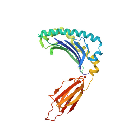
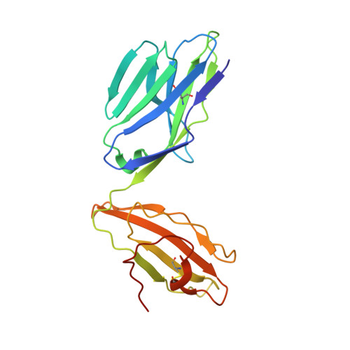
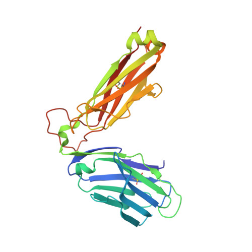
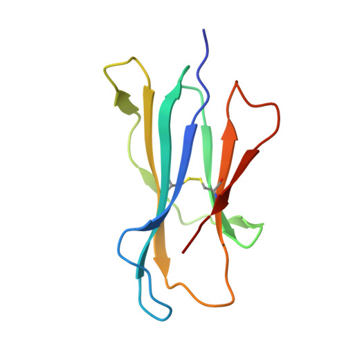
    The major-histocompatibility-complex-(MHC)-class-I-related molecule MR1 can present activating and non-activating vitamin-B-based ligands to mucosal-associated invariant T cells (MAIT cells). Whether MR1 binds other ligands is unknown. Here we identified a range of small organic molecules, drugs, drug metabolites and drug-like molecules, including salicylates and diclofenac, as MR1-binding ligands. Some of these ligands inhibited MAIT cells ex vivo and in vivo, while others, including diclofenac metabolites, were agonists. Crystal structures of a T cell antigen receptor (TCR) from a MAIT cell in complex with MR1 bound to the non-stimulatory and stimulatory compounds showed distinct ligand orientations and contacts within MR1, which highlighted the versatility of the MR1 binding pocket. The findings demonstrated that MR1 was able to capture chemically diverse structures, spanning mono- and bicyclic compounds, that either inhibited or activated MAIT cells. This indicated that drugs and drug-like molecules can modulate MAIT cell function in mammals.


  • Organizational Affiliation
    • Infection and Immunity Program and Department of Biochemistry and Molecular Biology, Biomedicine Discovery Institute, Monash University, Clayton, Australia.

Macromolecule Content 

  • Total Structure Weight: 188.67 kDa 
  • Atom Count: 13,060 
  • Modeled Residue Count: 1,605 
  • Deposited Residue Count: 1,638 
  • Unique protein chains: 4

Macromolecules

Find similar proteins by:|  3D Structure
Entity ID: 1
MoleculeChains  Sequence LengthOrganismDetailsImage
Major histocompatibility complex class I-related gene proteinA [auth C],
E [auth A]
271Homo sapiensMutation(s): 0 
Gene Names: MR1
UniProt & NIH Common Fund Data Resources
Find proteins for Q95460 (Homo sapiens)
Explore Q95460 
Go to UniProtKB:  Q95460
PHAROS:  Q95460
GTEx:  ENSG00000153029 
Entity Groups
Sequence Clusters30% Identity50% Identity70% Identity90% Identity95% Identity100% Identity
UniProt GroupQ95460
Sequence Annotations
Expand
Reference Sequence
Find similar proteins by:|  3D Structure
Entity ID: 2
MoleculeChains  Sequence LengthOrganismDetailsImage
MAIT T-cell receptor alpha chainB [auth D],
F [auth B]
203Homo sapiensMutation(s): 0 
Gene Names: B2M
Find similar proteins by:|  3D Structure
Entity ID: 3
MoleculeChains  Sequence LengthOrganismDetailsImage
MAIT T-cell receptor beta chainC [auth E],
G
245Homo sapiensMutation(s): 0 
Gene Names: TRAV/TRAC
Find similar proteins by:|  3D Structure
Entity ID: 4
MoleculeChains  Sequence LengthOrganismDetailsImage
Beta-2-microglobulinD [auth F],
H
100Homo sapiensMutation(s): 0 
Gene Names: B2MCDABP0092HDCMA22P
UniProt & NIH Common Fund Data Resources
Find proteins for P61769 (Homo sapiens)
Explore P61769 
Go to UniProtKB:  P61769
PHAROS:  P61769
GTEx:  ENSG00000166710 
Entity Groups
Sequence Clusters30% Identity50% Identity70% Identity90% Identity95% Identity100% Identity
UniProt GroupP61769
Sequence Annotations
Expand
Reference Sequence

Small Molecules

Ligands 4 Unique
IDChains Name / Formula / InChI Key2D Diagram3D Interactions
DIF

Query on DIF


Download:Ideal Coordinates CCD File
M [auth C],
Q [auth A]
2-[2,6-DICHLOROPHENYL)AMINO]BENZENEACETIC ACID
C14 H11 Cl2 N O2
DCOPUUMXTXDBNB-UHFFFAOYSA-N
GOL

Query on GOL


Download:Ideal Coordinates CCD File
K [auth C],
L [auth C],
P [auth A]
GLYCEROL
C3 H8 O3
PEDCQBHIVMGVHV-UHFFFAOYSA-N
ACT

Query on ACT


Download:Ideal Coordinates CCD File
J [auth C],
O [auth A],
R [auth B]
ACETATE ION
C2 H3 O2
QTBSBXVTEAMEQO-UHFFFAOYSA-M
NA

Query on NA


Download:Ideal Coordinates CCD File
I [auth C],
N [auth E]
SODIUM ION
Na
FKNQFGJONOIPTF-UHFFFAOYSA-N

Experimental Data & Validation

Experimental Data

  • Method: X-RAY DIFFRACTION
  • Resolution: 2.70 Å
  • R-Value Free:  0.237 (Depositor), 0.237 (DCC) 
  • R-Value Work:  0.187 (Depositor), 0.188 (DCC) 
  • R-Value Observed: 0.190 (Depositor) 
Space Group: C 1 2 1
Unit Cell:
Length ( Å )Angle ( ˚ )
a = 212.571α = 90
b = 69.65β = 103.38
c = 142.85γ = 90
Software Package:
Software NamePurpose
PHENIXrefinement
SCALAdata scaling
PHASERphasing
PDB_EXTRACTdata extraction
MOSFLMdata reduction

Structure Validation

View Full Validation Report


Ligand Structure Quality Assessment 


Entry History 

& Funding Information

Deposition Data


Funding OrganizationLocationGrant Number
Australian Research Council (ARC)AustraliaCE140100011
Australian Research Council (ARC)AustraliaAF50

Revision History  (Full details and data files)

  • Version 1.0: 2017-03-15
    Type: Initial release
  • Version 1.1: 2017-04-05
    Changes: Database references, Derived calculations
  • Version 1.2: 2020-01-01
    Changes: Author supporting evidence, Data collection
  • Version 1.3: 2023-10-04
    Changes: Data collection, Database references, Refinement description
  • Version 1.4: 2024-11-13
    Changes: Structure summary