5TZD | pdb_00005tzd

Structure of the WT S. venezulae BldD-(CTD-c-di-GMP)2 assembly intermediate


Experimental Data Snapshot

  • Method: X-RAY DIFFRACTION
  • Resolution: 1.75 Å
  • R-Value Free: 
    0.240 (Depositor), 0.244 (DCC) 
  • R-Value Work: 
    0.207 (Depositor), 0.213 (DCC) 
  • R-Value Observed: 
    0.215 (Depositor) 

Starting Model: experimental
View more details

wwPDB Validation 3D Report Full Report

Validation slider image for 5TZD

Ligand Structure Quality Assessment 


This is version 1.3 of the entry. See complete history

Literature

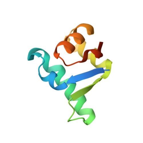
The Streptomyces master regulator BldD binds c-di-GMP sequentially to create a functional BldD2-(c-di-GMP)4 complex.

Schumacher, M.A.Zeng, W.Findlay, K.C.Buttner, M.J.Brennan, R.G.Tschowri, N.

(2017) Nucleic Acids Res 45: 6923-6933

  • DOI: https://doi.org/10.1093/nar/gkx287
  • Primary Citation Related Structures: 
    5TZD, 5TZF, 5TZG

  • PubMed Abstract: 

    Streptomyces are ubiquitous soil bacteria that undergo a complex developmental transition coinciding with their production of antibiotics. This transition is controlled by binding of a novel tetrameric form of the second messenger, 3΄-5΄ cyclic diguanylic acid (c-di-GMP) to the master repressor, BldD. In all domains of life, nucleotide-based second messengers allow a rapid integration of external and internal signals into regulatory pathways that control cellular responses to changing conditions. c-di-GMP can assume alternative oligomeric states to effect different functions, binding to effector proteins as monomers, intercalated dimers or, uniquely in the case of BldD, as a tetramer. However, at physiological concentrations c-di-GMP is a monomer and little is known about how higher oligomeric complexes assemble on effector proteins and if intermediates in assembly pathways have regulatory significance. Here, we show that c-di-GMP binds BldD using an ordered, sequential mechanism and that BldD function necessitates the assembly of the BldD2-(c-di-GMP)4 complex.


  • Organizational Affiliation
    • Department of Biochemistry, Duke University School of Medicine, Durham, NC 27701, USA.

Macromolecule Content 

  • Total Structure Weight: 43.46 kDa 
  • Atom Count: 2,828 
  • Modeled Residue Count: 296 
  • Deposited Residue Count: 364 
  • Unique protein chains: 1

Macromolecules

Find similar proteins by:|  3D Structure
Entity ID: 1
MoleculeChains  Sequence LengthOrganismDetailsImage
DNA-binding proteinA [auth J],
B,
C,
D [auth A]
91Streptomyces venezuelae ATCC 10712Mutation(s): 1 
Gene Names: SVEN_1089
UniProt
Find proteins for F2RCL8 (Streptomyces venezuelae (strain ATCC 10712 / CBS 650.69 / DSM 40230 / JCM 4526 / NBRC 13096 / PD 04745))
Explore F2RCL8 
Go to UniProtKB:  F2RCL8
Entity Groups
Sequence Clusters30% Identity50% Identity70% Identity90% Identity95% Identity100% Identity
UniProt GroupF2RCL8
Sequence Annotations
Expand
Reference Sequence

Small Molecules

Ligands 1 Unique
IDChains Name / Formula / InChI Key2D Diagram3D Interactions
C2E

Query on C2E


Download:Ideal Coordinates CCD File
E [auth J],
F [auth J],
G [auth C],
H [auth C]
9,9'-[(2R,3R,3aS,5S,7aR,9R,10R,10aS,12S,14aR)-3,5,10,12-tetrahydroxy-5,12-dioxidooctahydro-2H,7H-difuro[3,2-d:3',2'-j][1,3,7,9,2,8]tetraoxadiphosphacyclododecine-2,9-diyl]bis(2-amino-1,9-dihydro-6H-purin-6-one)
C20 H24 N10 O14 P2
PKFDLKSEZWEFGL-MHARETSRSA-N

Experimental Data & Validation

Experimental Data

  • Method: X-RAY DIFFRACTION
  • Resolution: 1.75 Å
  • R-Value Free:  0.240 (Depositor), 0.244 (DCC) 
  • R-Value Work:  0.207 (Depositor), 0.213 (DCC) 
  • R-Value Observed: 0.215 (Depositor) 
Space Group: P 61
Unit Cell:
Length ( Å )Angle ( ˚ )
a = 108.3α = 90
b = 108.3β = 90
c = 49.66γ = 120
Software Package:
Software NamePurpose
SCALAdata scaling
PHASERphasing
PHENIXrefinement
PDB_EXTRACTdata extraction
MOSFLMdata reduction

Structure Validation

View Full Validation Report


Ligand Structure Quality Assessment 


Entry History 

Deposition Data

Revision History  (Full details and data files)

  • Version 1.0: 2017-04-19
    Type: Initial release
  • Version 1.1: 2017-05-10
    Changes: Database references
  • Version 1.2: 2017-09-13
    Changes: Database references
  • Version 1.3: 2023-10-04
    Changes: Data collection, Database references, Refinement description, Structure summary