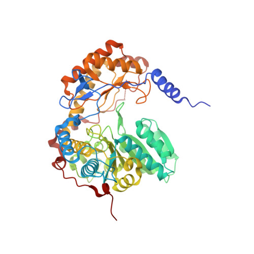
5TXR

Structure of ALAS from S. cerevisiae non-covalently bound to PLP cofactor

  • Classification: TRANSFERASE
  • Organism(s): Saccharomyces cerevisiae S288C
  • Expression System: Escherichia coli
  • Mutation(s): No 

  • Deposited: 2016-11-17 Released: 2018-03-28 
  • Deposition Author(s): Brown, B.L., Grant, R.A., Kardon, J.R., Sauer, R.T., Baker, T.A.
  • Funding Organization(s): Howard Hughes Medical Institute (HHMI), Burroughs Wellcome Fund, Jane Coffin Childs Memorial Fund for Medical Research, National Institutes of Health/National Institute Of Allergy and Infectious Diseases (NIH/NIAID), National Institutes of Health/National Institute of Diabetes and Digestive and Kidney Disease (NIH/NIDDK)

Experimental Data Snapshot

  • Method: X-RAY DIFFRACTION
  • Resolution: 1.90 Å
  • R-Value Free: 0.172 
  • R-Value Work: 0.142 
  • R-Value Observed: 0.142 

Starting Model: experimental
View more details

wwPDB Validation   3D Report Full Report


Ligand Structure Quality Assessment 


This is version 1.5 of the entry. See complete history


Literature

Structure of the Mitochondrial Aminolevulinic Acid Synthase, a Key Heme Biosynthetic Enzyme.

Brown, B.L.Kardon, J.R.Sauer, R.T.Baker, T.A.

(2018) Structure 26: 580-589.e4

  • DOI: https://doi.org/10.1016/j.str.2018.02.012
  • Primary Citation of Related Structures:  
    5TXR, 5TXT

  • PubMed Abstract: 

    5-Aminolevulinic acid synthase (ALAS) catalyzes the first step in heme biosynthesis. We present the crystal structure of a eukaryotic ALAS from Saccharomyces cerevisiae. In this homodimeric structure, one ALAS subunit contains covalently bound cofactor, pyridoxal 5'-phosphate (PLP), whereas the second is PLP free. Comparison between the subunits reveals PLP-coupled reordering of the active site and of additional regions to achieve the active conformation of the enzyme. The eukaryotic C-terminal extension, a region altered in multiple human disease alleles, wraps around the dimer and contacts active-site-proximal residues. Mutational analysis demonstrates that this C-terminal region that engages the active site is important for ALAS activity. Our discovery of structural elements that change conformation upon PLP binding and of direct contact between the C-terminal extension and the active site thus provides a structural basis for investigation of disruptions in the first step of heme biosynthesis and resulting human disorders.


  • Organizational Affiliation

    Department of Biology, Massachusetts Institute of Technology, Cambridge, MA 02139, USA.


Macromolecules
Find similar proteins by:  (by identity cutoff)  |  3D Structure
Entity ID: 1
MoleculeChains Sequence LengthOrganismDetailsImage
5-aminolevulinate synthase, mitochondrialA [auth B],
B [auth A]
491Saccharomyces cerevisiae S288CMutation(s): 0 
Gene Names: HEM1CYD1YDR232WYD9934.16
EC: 2.3.1.37
UniProt
Find proteins for P09950 (Saccharomyces cerevisiae (strain ATCC 204508 / S288c))
Explore P09950 
Go to UniProtKB:  P09950
Entity Groups  
Sequence Clusters30% Identity50% Identity70% Identity90% Identity95% Identity100% Identity
UniProt GroupP09950
Sequence Annotations
Expand
  • Reference Sequence
Small Molecules
Ligands 4 Unique
IDChains Name / Formula / InChI Key2D Diagram3D Interactions
PLP
Query on PLP

Download Ideal Coordinates CCD File 
G [auth B],
P [auth A]
PYRIDOXAL-5'-PHOSPHATE
C8 H10 N O6 P
NGVDGCNFYWLIFO-UHFFFAOYSA-N
GOL
Query on GOL

Download Ideal Coordinates CCD File 
H [auth A]GLYCEROL
C3 H8 O3
PEDCQBHIVMGVHV-UHFFFAOYSA-N
FMT
Query on FMT

Download Ideal Coordinates CCD File 
C [auth B]
D [auth B]
E [auth B]
F [auth B]
J [auth A]
C [auth B],
D [auth B],
E [auth B],
F [auth B],
J [auth A],
K [auth A],
L [auth A],
M [auth A],
N [auth A],
O [auth A]
FORMIC ACID
C H2 O2
BDAGIHXWWSANSR-UHFFFAOYSA-N
K
Query on K

Download Ideal Coordinates CCD File 
I [auth A]POTASSIUM ION
K
NPYPAHLBTDXSSS-UHFFFAOYSA-N
Experimental Data & Validation

Experimental Data

  • Method: X-RAY DIFFRACTION
  • Resolution: 1.90 Å
  • R-Value Free: 0.172 
  • R-Value Work: 0.142 
  • R-Value Observed: 0.142 
  • Space Group: P 1 21 1
Unit Cell:
Length ( Å )Angle ( ˚ )
a = 78.613α = 90
b = 64.878β = 95.86
c = 95.693γ = 90
Software Package:
Software NamePurpose
PHENIXrefinement
HKL-2000data reduction
HKL-2000data scaling
PHENIXphasing

Structure Validation

View Full Validation Report


Ligand Structure Quality Assessment 


Entry History & Funding Information

Deposition Data


Funding OrganizationLocationGrant Number
Howard Hughes Medical Institute (HHMI)United States--
Burroughs Wellcome FundUnited States1015092
Jane Coffin Childs Memorial Fund for Medical ResearchUnited States61-1529
National Institutes of Health/National Institute Of Allergy and Infectious Diseases (NIH/NIAID)United StatesAI-16892
National Institutes of Health/National Institute of Diabetes and Digestive and Kidney Disease (NIH/NIDDK)United StatesF32DK095726

Revision History  (Full details and data files)

  • Version 1.0: 2018-03-28
    Type: Initial release
  • Version 1.1: 2018-04-11
    Changes: Data collection, Database references
  • Version 1.2: 2018-04-18
    Changes: Data collection, Database references
  • Version 1.3: 2019-11-20
    Changes: Author supporting evidence
  • Version 1.4: 2024-03-06
    Changes: Data collection, Database references
  • Version 1.5: 2024-04-03
    Changes: Refinement description