5TTH

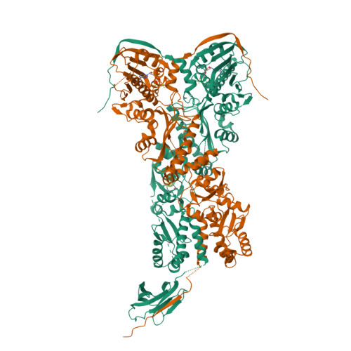
Heterodimeric SpyCatcher/SpyTag-fused zebrafish TRAP1 in ATP/ADP-hybrid state


Experimental Data Snapshot

  • Method: X-RAY DIFFRACTION
  • Resolution: 3.20 Å
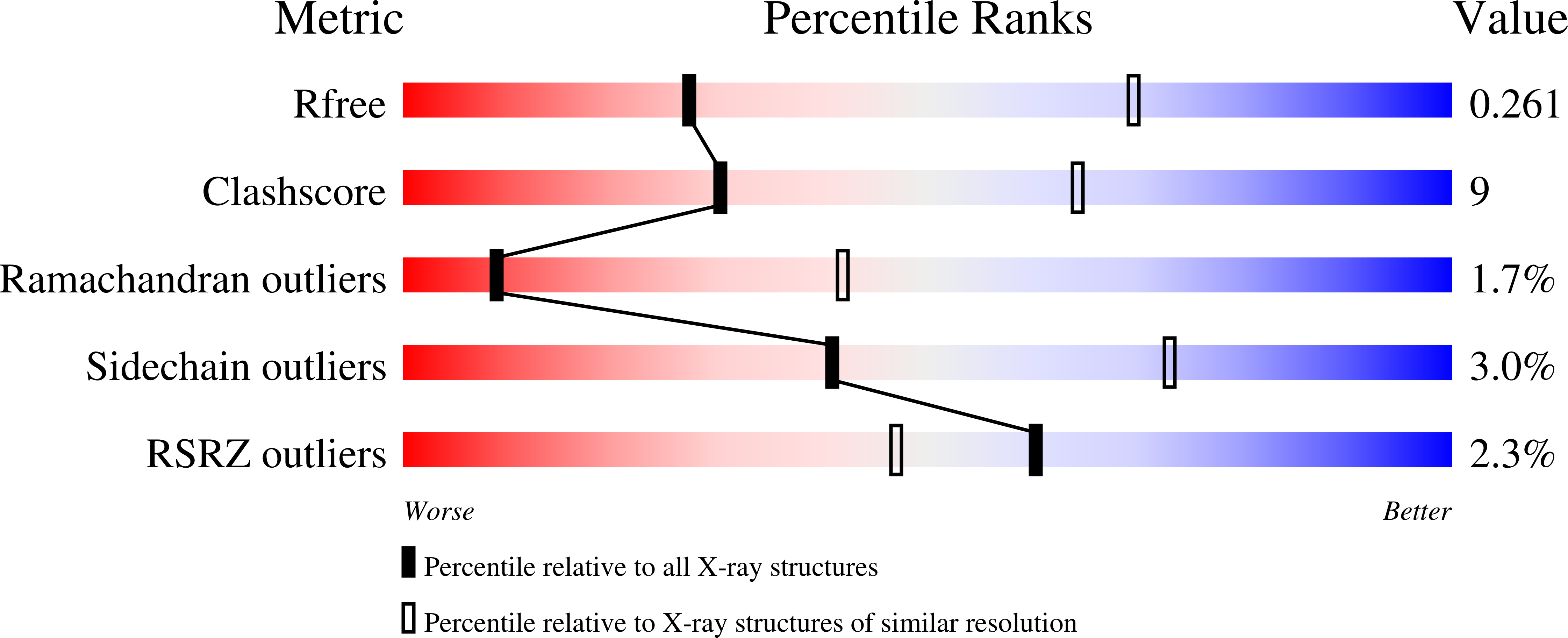
  • R-Value Free: 0.261 
  • R-Value Work: 0.228 
  • R-Value Observed: 0.230 

Starting Model: experimental
View more details

wwPDB Validation   3D Report Full Report


Ligand Structure Quality Assessment 


This is version 1.5 of the entry. See complete history


Literature

Symmetry broken and rebroken during the ATP hydrolysis cycle of the mitochondrial Hsp90 TRAP1.

Elnatan, D.Betegon, M.Liu, Y.Ramelot, T.Kennedy, M.A.Agard, D.A.

(2017) Elife 6

  • DOI: https://doi.org/10.7554/eLife.25235
  • Primary Citation of Related Structures:  
    5TTH, 5TVU, 5TVW, 5TVX

  • PubMed Abstract: 

    Hsp90 is a homodimeric ATP-dependent molecular chaperone that remodels its substrate 'client' proteins, facilitating their folding and activating them for biological function. Despite decades of research, the mechanism connecting ATP hydrolysis and chaperone function remains elusive. Particularly puzzling has been the apparent lack of cooperativity in hydrolysis of the ATP in each protomer. A crystal structure of the mitochondrial Hsp90, TRAP1, revealed that the catalytically active state is closed in a highly strained asymmetric conformation. This asymmetry, unobserved in other Hsp90 homologs, is due to buckling of one of the protomers and is most pronounced at the broadly conserved client-binding region. Here, we show that rather than being cooperative or independent, ATP hydrolysis on the two protomers is sequential and deterministic. Moreover, dimer asymmetry sets up differential hydrolysis rates for each protomer, such that the buckled conformation favors ATP hydrolysis. Remarkably, after the first hydrolysis, the dimer undergoes a flip in the asymmetry while remaining in a closed state for the second hydrolysis. From these results, we propose a model where direct coupling of ATP hydrolysis and conformational flipping rearranges client-binding sites, providing a paradigm of how energy from ATP hydrolysis can be used for client remodeling.


  • Organizational Affiliation

    Department of Biochemistry and Biophysics, Howard Hughes Medical Institute, University of California, San Francisco, United States.


Macromolecules
Find similar proteins by:  (by identity cutoff)  |  3D Structure
Entity ID: 1
MoleculeChains Sequence LengthOrganismDetailsImage
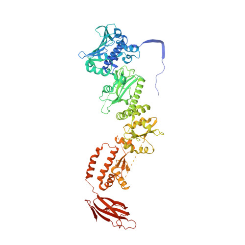
C-terminal SpyCatcher fusion of wildtype zebrafish TNF receptor-associated protein 1738Danio rerioStreptococcus pyogenes
This entity is chimeric
Mutation(s): 2 
Gene Names: trap1fba2
UniProt
Find proteins for A8WFV1 (Danio rerio)
Explore A8WFV1 
Go to UniProtKB:  A8WFV1
Find proteins for Q8G9G1 (Streptococcus pyogenes)
Explore Q8G9G1 
Go to UniProtKB:  Q8G9G1
Entity Groups  
Sequence Clusters30% Identity50% Identity70% Identity90% Identity95% Identity100% Identity
UniProt GroupsQ8G9G1A8WFV1
Sequence Annotations
Expand
  • Reference Sequence
Find similar proteins by:  (by identity cutoff)  |  3D Structure
Entity ID: 2
MoleculeChains Sequence LengthOrganismDetailsImage
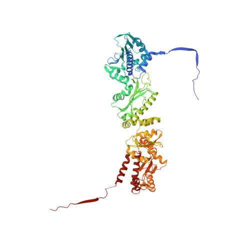
C-terminal Spytag fusion of R417A zebrafish TNF receptor-associated protein 1672Danio rerioStreptococcus pyogenes
This entity is chimeric
Mutation(s): 1 
Gene Names: trap1fba2
UniProt
Find proteins for A8WFV1 (Danio rerio)
Explore A8WFV1 
Go to UniProtKB:  A8WFV1
Find proteins for Q8G9G1 (Streptococcus pyogenes)
Explore Q8G9G1 
Go to UniProtKB:  Q8G9G1
Entity Groups  
Sequence Clusters30% Identity50% Identity70% Identity90% Identity95% Identity100% Identity
UniProt GroupsQ8G9G1A8WFV1
Sequence Annotations
Expand
  • Reference Sequence
Experimental Data & Validation

Experimental Data

  • Method: X-RAY DIFFRACTION
  • Resolution: 3.20 Å
  • R-Value Free: 0.261 
  • R-Value Work: 0.228 
  • R-Value Observed: 0.230 
  • Space Group: C 1 2 1
Unit Cell:
Length ( Å )Angle ( ˚ )
a = 180.086α = 90
b = 95.711β = 134.59
c = 126.558γ = 90
Software Package:
Software NamePurpose
REFMACrefinement
PDB_EXTRACTdata extraction
XDSdata reduction
PHASERphasing

Structure Validation

View Full Validation Report


Ligand Structure Quality Assessment 


Entry History & Funding Information

Deposition Data


Funding OrganizationLocationGrant Number
National Institutes of Health/National Institute of General Medical Sciences (NIH/NIGMS)United StatesU01 GM098254.

Revision History  (Full details and data files)

  • Version 1.0: 2017-08-23
    Type: Initial release
  • Version 1.1: 2017-09-20
    Changes: Author supporting evidence
  • Version 1.2: 2019-05-08
    Changes: Data collection, Database references
  • Version 1.3: 2020-01-01
    Changes: Author supporting evidence
  • Version 1.4: 2023-10-04
    Changes: Data collection, Database references, Derived calculations, Refinement description
  • Version 1.5: 2024-10-16
    Changes: Structure summary