5TT5 | pdb_00005tt5

Escherichia coli LigA (K115M) in complex with NAD+


Experimental Data Snapshot

  • Method: X-RAY DIFFRACTION
  • Resolution: 1.55 Å
  • R-Value Free: 
    0.205 (Depositor), 0.205 (DCC) 
  • R-Value Work: 
    0.176 (Depositor), 0.178 (DCC) 
  • R-Value Observed: 
    0.178 (Depositor) 

Starting Model: experimental
View more details

wwPDB Validation 3D Report Full Report

Validation slider image for 5TT5

Ligand Structure Quality Assessment 


This is version 1.4 of the entry. See complete history

Literature

Two-metal versus one-metal mechanisms of lysine adenylylation by ATP-dependent and NAD(+)-dependent polynucleotide ligases.

Unciuleac, M.C.Goldgur, Y.Shuman, S.

(2017) Proc Natl Acad Sci U S A 114: 2592-2597

  • DOI: https://doi.org/10.1073/pnas.1619220114
  • Primary Citation Related Structures: 
    5TT5, 5TT6

  • PubMed Abstract: 

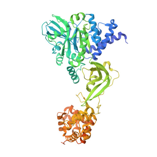
    Polynucleotide ligases comprise a ubiquitous superfamily of nucleic acid repair enzymes that join 3'-OH and 5'-PO 4 DNA or RNA ends. Ligases react with ATP or NAD + and a divalent cation cofactor to form a covalent enzyme-(lysine-Nζ)-adenylate intermediate. Here, we report crystal structures of the founding members of the ATP-dependent RNA ligase family (T4 RNA ligase 1; Rnl1) and the NAD + -dependent DNA ligase family ( Escherichia coli LigA), captured as their respective Michaelis complexes, which illuminate distinctive catalytic mechanisms of the lysine adenylylation reaction. The 2.2-Å Rnl1•ATP•(Mg 2+ ) 2 structure highlights a two-metal mechanism, whereby: a ligase-bound "catalytic" Mg 2+ (H 2 O) 5 coordination complex lowers the p K a of the lysine nucleophile and stabilizes the transition state of the ATP α phosphate; a second octahedral Mg 2+ coordination complex bridges the β and γ phosphates; and protein elements unique to Rnl1 engage the γ phosphate and associated metal complex and orient the pyrophosphate leaving group for in-line catalysis. By contrast, the 1.55-Å LigA•NAD + •Mg 2+ structure reveals a one-metal mechanism in which a ligase-bound Mg 2+ (H 2 O) 5 complex lowers the lysine p K a and engages the NAD + α phosphate, but the β phosphate and the nicotinamide nucleoside of the nicotinamide mononucleotide (NMN) leaving group are oriented solely via atomic interactions with protein elements that are unique to the LigA clade. The two-metal versus one-metal dichotomy demarcates a branchpoint in ligase evolution and favors LigA as an antibacterial drug target.


  • Organizational Affiliation
    • Molecular Biology Program, Sloan-Kettering Institute, New York, NY 10065.

Macromolecule Content 

  • Total Structure Weight: 76.93 kDa 
  • Atom Count: 5,407 
  • Modeled Residue Count: 581 
  • Deposited Residue Count: 691 
  • Unique protein chains: 1

Macromolecules

Find similar proteins by:|  3D Structure
Entity ID: 1
MoleculeChains  Sequence LengthOrganismDetailsImage
DNA ligase691Escherichia coli K-12Mutation(s): 1 
Gene Names: ligAdnaLligloppdeCb2411JW2403
EC: 6.5.1.2
UniProt
Find proteins for P15042 (Escherichia coli (strain K12))
Explore P15042 
Go to UniProtKB:  P15042
Entity Groups
Sequence Clusters30% Identity50% Identity70% Identity90% Identity95% Identity100% Identity
UniProt GroupP15042
Sequence Annotations
Expand
Reference Sequence

Experimental Data & Validation

Experimental Data

  • Method: X-RAY DIFFRACTION
  • Resolution: 1.55 Å
  • R-Value Free:  0.205 (Depositor), 0.205 (DCC) 
  • R-Value Work:  0.176 (Depositor), 0.178 (DCC) 
  • R-Value Observed: 0.178 (Depositor) 
Space Group: P 21 21 21
Unit Cell:
Length ( Å )Angle ( ˚ )
a = 40.907α = 90
b = 94.312β = 90
c = 150.927γ = 90
Software Package:
Software NamePurpose
PHENIXrefinement
HKL-2000data reduction
HKL-2000data scaling
MOLREPphasing

Structure Validation

View Full Validation Report


Ligand Structure Quality Assessment 


Entry History 

& Funding Information

Deposition Data


Funding OrganizationLocationGrant Number
National Institutes of Health/National Institute of General Medical Sciences (NIH/NIGMS)United StatesGM63611-14
National Institutes of Health/National Cancer Institute (NIH/NCI)United StatesP30-CA008748

Revision History  (Full details and data files)

  • Version 1.0: 2017-03-08
    Type: Initial release
  • Version 1.1: 2017-03-22
    Changes: Database references
  • Version 1.2: 2017-09-20
    Changes: Author supporting evidence
  • Version 1.3: 2019-12-04
    Changes: Author supporting evidence
  • Version 1.4: 2023-10-04
    Changes: Data collection, Database references, Derived calculations, Refinement description